4RRT image
Entry Detail
PDB ID:
4RRT
Keywords:
Title:
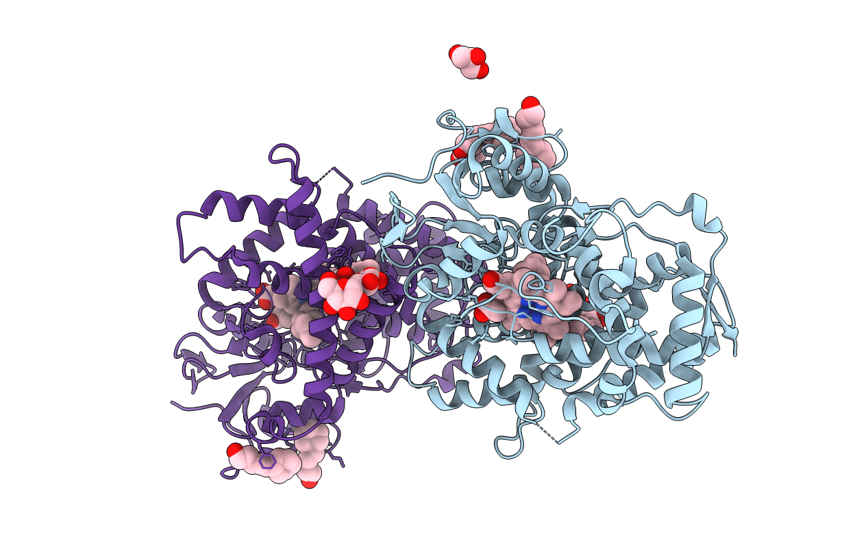
Crystal structure of a human cytochrome P450 2B6 (Y226H/K262R) in complex with (+)-3-carene
Biological Source:
Source Organism:
Host Organism:
PDB Version:
Deposition Date:
2014-11-06
Release Date:
2015-01-28
Method Details:
Experimental Method:
Resolution:
2.20 Å
R-Value Free:
0.22
R-Value Work:
0.17
R-Value Observed:
0.17
Space Group:
P 32
Macromolecular Entities
Polymer Type:polypeptide(L)
Description:Cytochrome P450 2B6
Mutations:Y226H, K262R
Chain IDs:A, B
Chain Length:476
Number of Molecules:2
Biological Source:Homo sapiens
Primary Citation
Structural and Biophysical Characterization of Human Cytochromes P450 2B6 and 2A6 Bound to Volatile Hydrocarbons: Analysis and Comparison.
Mol.Pharmacol. 87 649 659 (2015)
PMID: 25585967 DOI: 10.1124/mol.114.097014

Abstact

X-ray crystal structures of complexes of cytochromes CYP2B6 and CYP2A6 with the monoterpene sabinene revealed two distinct binding modes in the active sites. In CYP2B6, sabinene positioned itself with the putative oxidation site located closer to the heme iron. In contrast, sabinene was found in an alternate conformation in the more compact CYP2A6, where the larger hydrophobic side chains resulted in a significantly reduced active-site cavity. Furthermore, results from isothermal titration calorimetry indicated a much more substantial contribution of favorable enthalpy to sabinene binding to CYP2B6 as opposed to CYP2A6, consistent with the previous observations with (+)-α-pinene. Structural analysis of CYP2B6 complexes with sabinene and the structurally similar (3)-carene and comparison with previously solved structures revealed how the movement of the F206 side chain influences the volume of the binding pocket. In addition, retrospective analysis of prior structures revealed that ligands containing -Cl and -NH functional groups adopted a distinct orientation in the CYP2B active site compared with other ligands. This binding mode may reflect the formation of Cl-π or NH-π bonds with aromatic rings in the active site, which serve as important contributors to protein-ligand binding affinity and specificity. Overall, the findings from multiple techniques illustrate how drugs metabolizing CYP2B6 and CYP2A6 handle a common hydrocarbon found in the environment. The study also provides insight into the role of specific functional groups of the ligand that may influence the binding to CYP2B6.

Legend

Protein

Chemical

Disease

Primary Citation of related structures