
Deposition Date
2014-10-30
Release Date
2015-01-21
Last Version Date
2023-09-20
Entry Detail
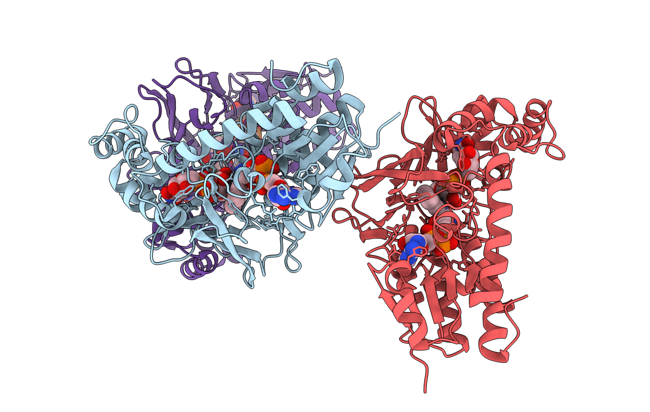
PDB ID:
4RPH
Keywords:
Title:
Crystal structure of Micobacterium tuberculosis UDP-Galactopyranose mutase in complex with substrate UDP-Galp (reduced)
Biological Source:
Source Organism(s):
Mycobacterium tuberculosis (Taxon ID: 83332)
Expression System(s):
Method Details:
Experimental Method:
Resolution:
2.60 Å
R-Value Free:
0.24
R-Value Work:
0.20
R-Value Observed:
0.20
Space Group:
C 1 2 1
