4RND image
Deposition Date 2014-10-24
Release Date 2014-12-10
Last Version Date 2024-02-28
Entry Detail
PDB ID:
4RND
Keywords:
Title:
Crystal Structure of the subunit DF-assembly of the eukaryotic V-ATPase.
Biological Source:
Source Organism:
Host Organism:
Method Details:
Experimental Method:
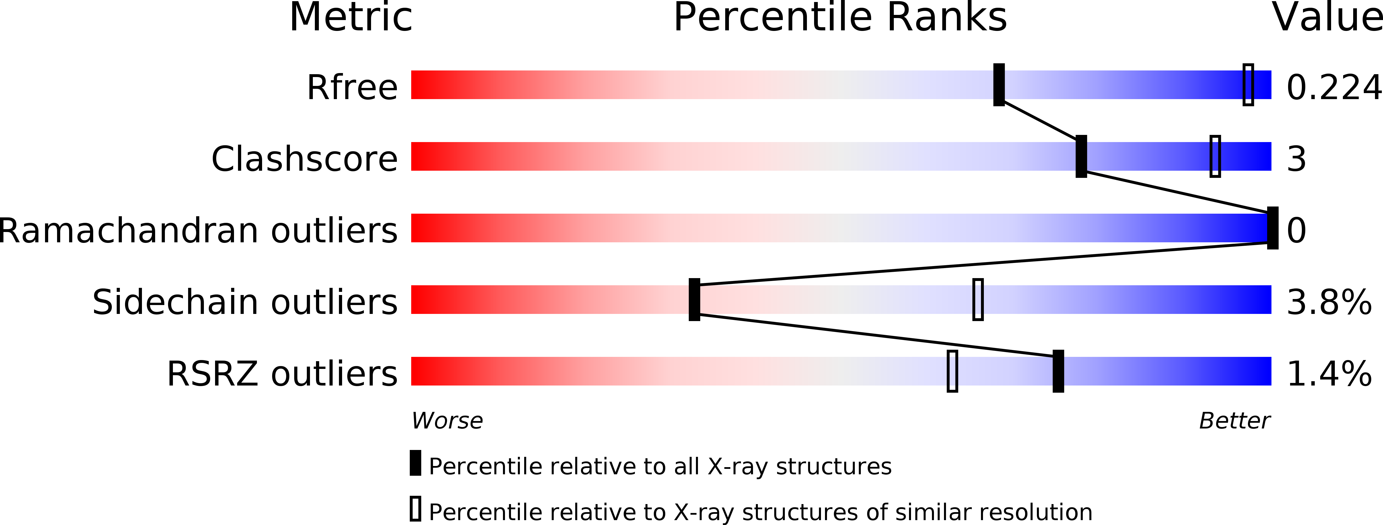
Resolution:
3.18 Å
R-Value Free:
0.23
R-Value Work:
0.20
R-Value Observed:
0.20
Space Group:
P 61
Macromolecular Entities
Structures with similar UniProt ID
Protein Blast
Polymer Type:polypeptide(L)
Molecule:V-type proton ATPase subunit D
Gene (Uniprot):VMA8
Chain IDs:A, C
Chain Length:256
Number of Molecules:2
Biological Source:Saccharomyces cerevisiae S288c
Structures with similar UniProt ID
Protein Blast
Polymer Type:polypeptide(L)
Molecule:V-type proton ATPase subunit F
Gene (Uniprot):VMA7
Chain IDs:B, D
Chain Length:118
Number of Molecules:2
Biological Source:Saccharomyces cerevisiae S288c
Ligand Molecules
Primary Citation
Crystal Structure of Subunits D and F in Complex Gives Insight into Energy Transmission of the Eukaryotic V-ATPase from Saccharomyces cerevisiae.
J.Biol.Chem. 290 3183 3196 (2015)
PMID: 25505269 DOI: 10.1074/jbc.M114.622688

Abstact

Eukaryotic V1VO-ATPases hydrolyze ATP in the V1 domain coupled to ion pumping in VO. A unique mode of regulation of V-ATPases is the reversible disassembly of V1 and VO, which reduces ATPase activity and causes silencing of ion conduction. The subunits D and F are proposed to be key in these enzymatic processes. Here, we describe the structures of two conformations of the subunit DF assembly of Saccharomyces cerevisiae (ScDF) V-ATPase at 3.1 Å resolution. Subunit D (ScD) consists of a long pair of α-helices connected by a short helix ((79)IGYQVQE(85)) as well as a β-hairpin region, which is flanked by two flexible loops. The long pair of helices is composed of the N-terminal α-helix and the C-terminal helix, showing structural alterations in the two ScDF structures. The entire subunit F (ScF) consists of an N-terminal domain of four β-strands (β1-β4) connected by four α-helices (α1-α4). α1 and β2 are connected via the loop (26)GQITPETQEK(35), which is unique in eukaryotic V-ATPases. Adjacent to the N-terminal domain is a flexible loop, followed by a C-terminal α-helix (α5). A perpendicular and extended conformation of helix α5 was observed in the two crystal structures and in solution x-ray scattering experiments, respectively. Fitted into the nucleotide-bound A3B3 structure of the related A-ATP synthase from Enterococcus hirae, the arrangements of the ScDF molecules reflect their central function in ATPase-coupled ion conduction. Furthermore, the flexibility of the terminal helices of both subunits as well as the loop (26)GQITPETQEK(35) provides information about the regulatory step of reversible V1VO disassembly.

Legend

Protein

Chemical

Disease

Primary Citation of related structures