4RMB image
Deposition Date 2014-10-21
Release Date 2015-02-11
Last Version Date 2023-09-20
Entry Detail
PDB ID:
4RMB
Keywords:
Title:
Crystal structure of keratin 4 binding domain of surface adhesin Srr-1 of S.agalactiae
Biological Source:
Source Organism:
Host Organism:
Method Details:
Experimental Method:
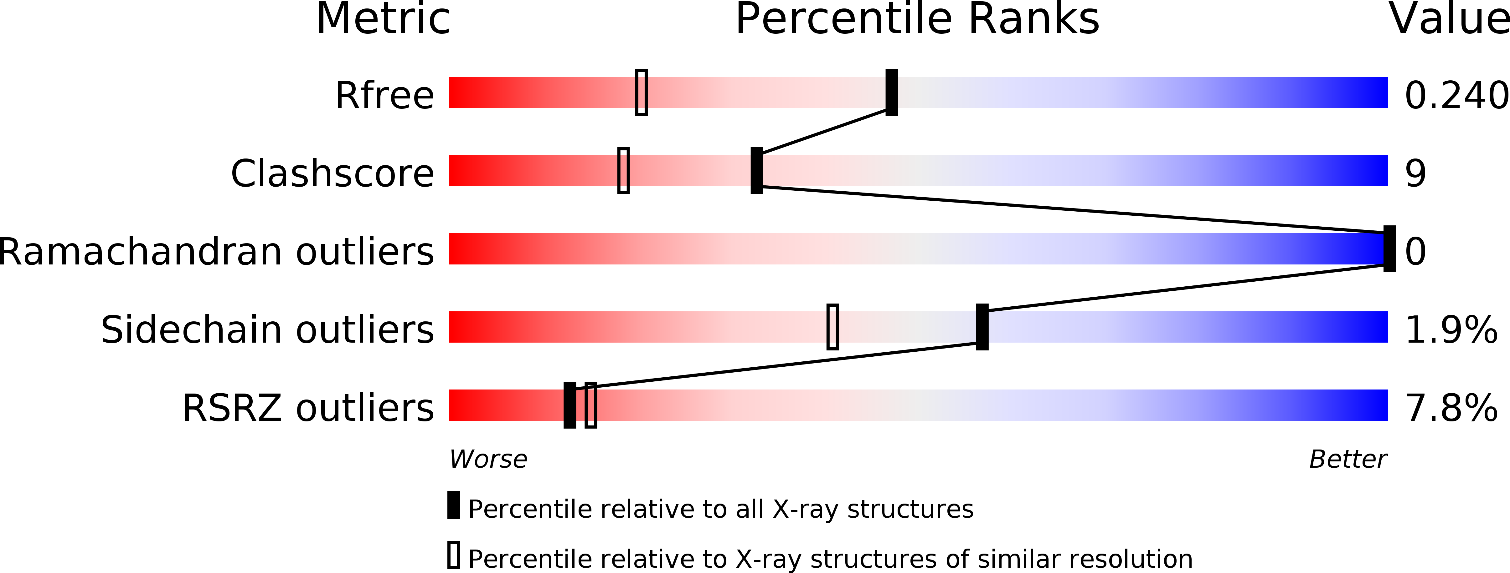
Resolution:
1.70 Å
R-Value Free:
0.24
R-Value Work:
0.22
Space Group:
P 1 21 1
Macromolecular Entities
Structures with similar UniProt ID
Protein Blast
Polymer Type:polypeptide(L)
Molecule:Serine rich repeat protein-1 (Srr-1)
Gene (Uniprot):gbs1529
Chain IDs:A, B
Chain Length:166
Number of Molecules:2
Biological Source:Streptococcus agalactiae NEM316
Primary Citation
Structure of KRT4 binding domain of Srr-1 from Streptococcus agalactiae reveals a novel beta-sheet complementation.
Int.J.Biol.Macromol. 75C 97 105 (2015)
PMID: 25603146 DOI: 10.1016/j.ijbiomac.2014.12.048

Abstact

The serine rich repeat protein-1 (Srr-1) is an adhesive protein of Streptococcus agalactiae. It is the first bacterial protein identified to interact with human keratin 4 (K4 or KRT4). Within Srr-1, the residues 311-641 constitute the non-repeat ligand binding region (Srr-1-BR(311-641)). The C-terminal part of Srr-1-BR(311-641), comprising of residues 485-642 (termed Srr-1-K4BD), have been identified to bind to K4. Here we report the crystal structure of recombinant Srr-1-K4BD(485-642) and its possible mode of interaction with K4 through docking studies. The dimeric structure of Srr-1-K4BD(485-642) reveals a novel two way "slide lock" parallel β-sheet complementation where the C-terminal strand of one monomer is positioned anti-parallel to the N-terminal strand of the adjacent monomer and this arrangement is not seen so far in any of the homologous structures. The dimerization of Srr-1-K4BD(485-642) observed both in the crystal structure and in solution suggests that similar domain association could also be possible in in vivo and we propose this association would likely generate a new binding site for another host molecule. It is likely that the adhesin can recognize multiple ligands using its ligand binding sub-domains through their intra and inter domain association with one another.

Legend

Protein

Chemical

Disease

Primary Citation of related structures