4RLY image
Deposition Date 2014-10-18
Release Date 2014-11-26
Last Version Date 2024-02-28
Entry Detail
PDB ID:
4RLY
Title:
Crystal Structure of AnkB Ankyrin Repeats (R1-R9) in Complex with Nav1.2 Ankyrin Binding Domain
Biological Source:
Source Organism:
Mus musculus (Taxon ID: 10090)
Homo sapiens (Taxon ID: 9606)
Host Organism:
Method Details:
Experimental Method:
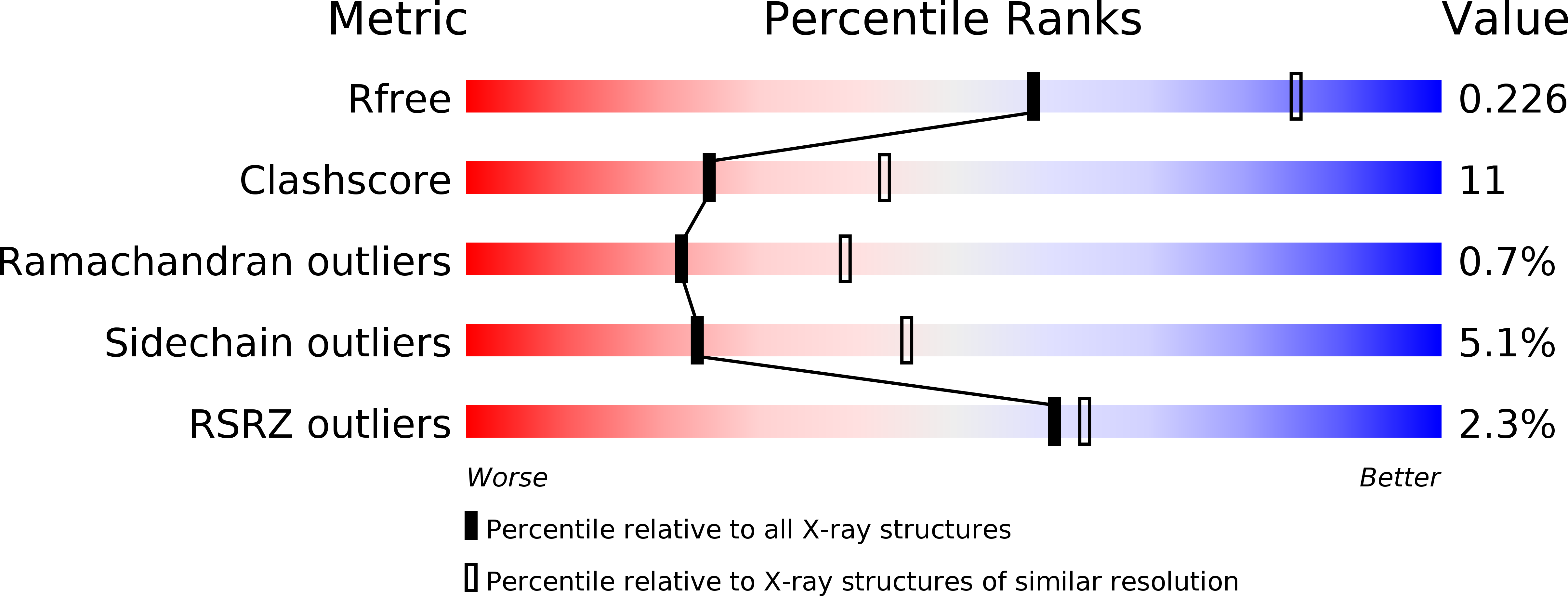
Resolution:
2.50 Å
R-Value Free:
0.23
R-Value Work:
0.18
R-Value Observed:
0.19
Space Group:
P 42 2 2
Macromolecular Entities
Protein Blast
Polymer Type:polypeptide(L)
Molecule:Nav1.2 - AnkB chimera
Gene (Uniprot):ANK2
Chain IDs:A
Chain Length:329
Number of Molecules:1
Biological Source:Mus musculus, Homo sapiens
Ligand Molecules
Primary Citation
Structural basis of diverse membrane target recognitions by ankyrins.
Elife 3 e04353 ? (2014)
PMID: 25383926 DOI: 10.7554/eLife.04353

Abstact

Ankyrin adaptors together with their spectrin partners coordinate diverse ion channels and cell adhesion molecules within plasma membrane domains and thereby promote physiological activities including fast signaling in the heart and nervous system. Ankyrins specifically bind to numerous membrane targets through their 24 ankyrin repeats (ANK repeats), although the mechanism for the facile and independent evolution of these interactions has not been resolved. Here we report the structures of ANK repeats in complex with an inhibitory segment from the C-terminal regulatory domain and with a sodium channel Nav1.2 peptide, respectively, showing that the extended, extremely conserved inner groove spanning the entire ANK repeat solenoid contains multiple target binding sites capable of accommodating target proteins with very diverse sequences via combinatorial usage of these sites. These structures establish a framework for understanding the evolution of ankyrins' membrane targets, with implications for other proteins containing extended ANK repeat domains.

Legend

Protein

Chemical

Disease

Primary Citation of related structures