4RI3 image
Deposition Date 2014-10-05
Release Date 2015-08-12
Last Version Date 2024-11-27
Entry Detail
PDB ID:
4RI3
Title:
Crystal structure of DCCD-modified PsbS from spinach
Biological Source:
Source Organism:
Method Details:
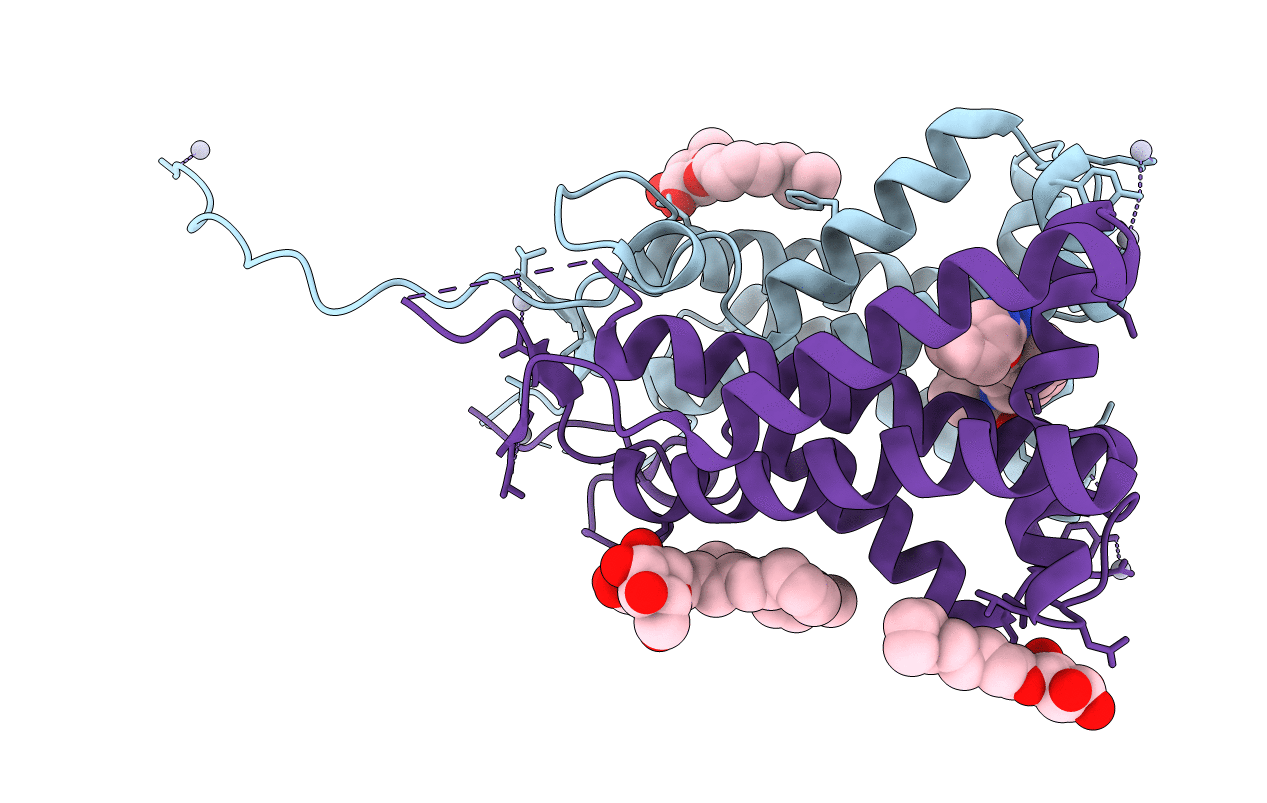
Experimental Method:
Resolution:
2.70 Å
R-Value Free:
0.28
R-Value Work:
0.23
R-Value Observed:
0.23
Space Group:
P 21 21 21
Macromolecular Entities
Structures with similar UniProt ID
Protein Blast
Polymer Type:polypeptide(L)
Molecule:Photosystem II 22 kDa protein, chloroplastic
Gene (Uniprot):PSBS
Chain IDs:A, B
Chain Length:212
Number of Molecules:2
Biological Source:Spinacia oleracea
Primary Citation
Crystal structures of the PsbS protein essential for photoprotection in plants.
Nat.Struct.Mol.Biol. 22 729 735 (2015)
PMID: 26258636 DOI: 10.1038/nsmb.3068

Abstact

The photosystem II protein PsbS has an essential role in qE-type nonphotochemical quenching, which protects plants from photodamage under excess light conditions. qE is initiated by activation of PsbS by low pH, but the mechanism of PsbS action remains elusive. Here we report the low-pH crystal structures of PsbS from spinach in its free form and in complex with the qE inhibitor N,N'-dicyclohexylcarbodiimide (DCCD), revealing that PsbS adopts a unique folding pattern, and, unlike other members of the light-harvesting-complex superfamily, it is a noncanonical pigment-binding protein. Structural and biochemical evidence shows that both active and inactive PsbS form homodimers in the thylakoid membranes, and DCCD binding disrupts the lumenal intermolecular hydrogen bonds of the active PsbS dimer. Activation of PsbS by low pH during qE may involve a conformational change associated with altered lumenal intermolecular interactions of the PsbS dimer.

Legend

Protein

Chemical

Disease

Primary Citation of related structures
Feedback Form
Name
Email
Institute
Feedback