4RH7 image
Deposition Date 2014-10-01
Release Date 2014-12-10
Last Version Date 2024-02-28
Entry Detail
PDB ID:
4RH7
Keywords:
Title:
Crystal structure of human cytoplasmic dynein 2 motor domain in complex with ADP.Vi
Biological Source:
Source Organism:
Host Organism:
Method Details:
Experimental Method:
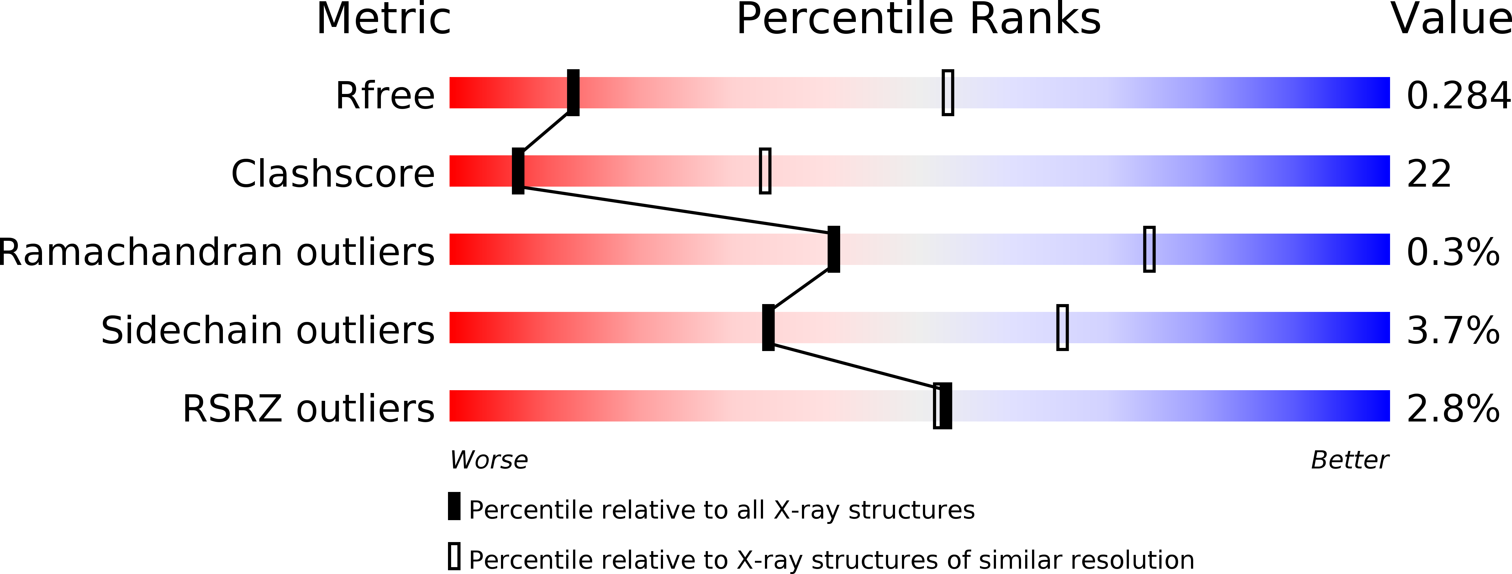
Resolution:
3.41 Å
R-Value Free:
0.28
R-Value Work:
0.23
R-Value Observed:
0.23
Space Group:
C 2 2 21
Macromolecular Entities
Structures with similar UniProt ID
Protein Blast
Polymer Type:polypeptide(L)
Molecule:Green fluorescent protein/Cytoplasmic dynein 2 heavy chain 1
Gene (Uniprot):DYNC2H1
Chain IDs:A
Chain Length:3450
Number of Molecules:1
Biological Source:synthetic construct, Homo sapiens
Primary Citation
Structure of human cytoplasmic dynein-2 primed for its power stroke.
Nature 518 435 438 (2015)
PMID: 25470043 DOI: 10.1038/nature14023

Abstact

Members of the dynein family, consisting of cytoplasmic and axonemal isoforms, are motors that move towards the minus ends of microtubules. Cytoplasmic dynein-1 (dynein-1) plays roles in mitosis and cellular cargo transport, and is implicated in viral infections and neurodegenerative diseases. Cytoplasmic dynein-2 (dynein-2) performs intraflagellar transport and is associated with human skeletal ciliopathies. Dyneins share a conserved motor domain that couples cycles of ATP hydrolysis with conformational changes to produce movement. Here we present the crystal structure of the human cytoplasmic dynein-2 motor bound to the ATP-hydrolysis transition state analogue ADP.vanadate. The structure reveals a closure of the motor's ring of six AAA+ domains (ATPases associated with various cellular activites: AAA1-AAA6). This induces a steric clash with the linker, the key element for the generation of movement, driving it into a conformation that is primed to produce force. Ring closure also changes the interface between the stalk and buttress coiled-coil extensions of the motor domain. This drives helix sliding in the stalk which causes the microtubule binding domain at its tip to release from the microtubule. Our structure answers the key questions of how ATP hydrolysis leads to linker remodelling and microtubule affinity regulation.

Legend

Protein

Chemical

Disease

Primary Citation of related structures