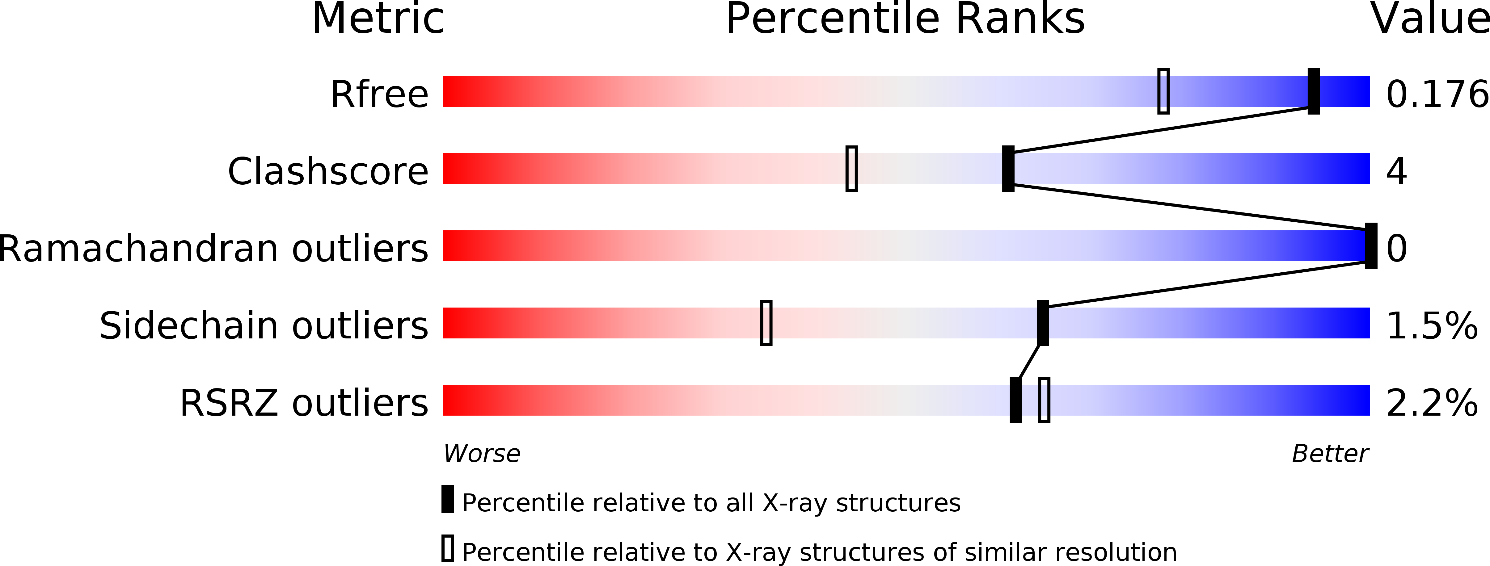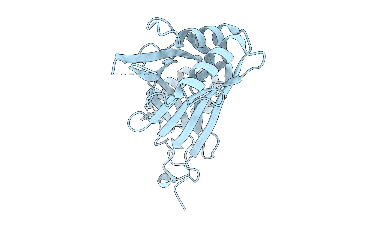
4RAX image
Deposition Date 2014-09-11
Release Date 2015-09-23
Last Version Date 2024-11-06
Entry Detail
PDB ID:
4RAX
Title:
A regulatory domain of an ion channel
Biological Source:
Source Organism:
Mus musculus (Taxon ID: 10090)
Host Organism:
Method Details:
Experimental Method:
Resolution:
1.45 Å
R-Value Free:
0.17
R-Value Work:
0.15
R-Value Observed:
0.15
Space Group:
P 21 3
Macromolecular Entities
Polymer Type:polypeptide(L)
Molecule:Piezo-type mechanosensitive ion channel component 1
Gene (Uniprot):Piezo1
Chain IDs:A
Chain Length:252
Number of Molecules:1
Biological Source:Mus musculus
Primary Citation
Architecture of the mammalian mechanosensitive Piezo1 channel.
Nature 527 64 69 (2015)
PMID: 26390154 DOI: 10.1038/nature15247

Abstact

Piezo proteins are evolutionarily conserved and functionally diverse mechanosensitive cation channels. However, the overall structural architecture and gating mechanisms of Piezo channels have remained unknown. Here we determine the cryo-electron microscopy structure of the full-length (2,547 amino acids) mouse Piezo1 (Piezo1) at a resolution of 4.8 Å. Piezo1 forms a trimeric propeller-like structure (about 900 kilodalton), with the extracellular domains resembling three distal blades and a central cap. The transmembrane region has 14 apparently resolved segments per subunit. These segments form three peripheral wings and a central pore module that encloses a potential ion-conducting pore. The rather flexible extracellular blade domains are connected to the central intracellular domain by three long beam-like structures. This trimeric architecture suggests that Piezo1 may use its peripheral regions as force sensors to gate the central ion-conducting pore.

Legend

Protein

Chemical

Disease

Primary Citation of related structures