Deposition Date
2014-09-08
Release Date
2014-12-03
Last Version Date
2023-09-20
Entry Detail
PDB ID:
4R9V
Keywords:
Title:
Crystal structure of sialyltransferase from photobacterium damselae, residues 113-497 corresponding to the gt-b domain
Biological Source:
Source Organism(s):
Photobacterium damselae (Taxon ID: 38293)
Expression System(s):
Method Details:
Experimental Method:
Resolution:
2.30 Å
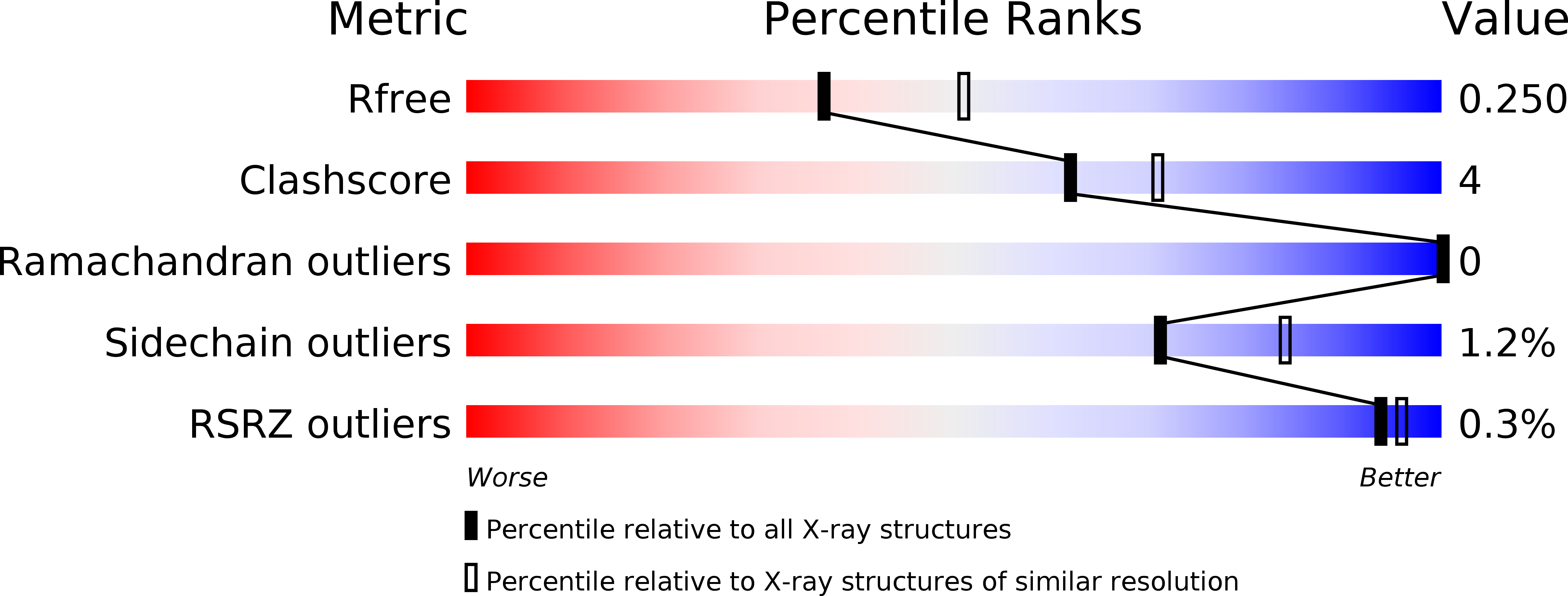
R-Value Free:
0.24
R-Value Work:
0.18
R-Value Observed:
0.18
Space Group:
C 1 2 1