Deposition Date
2014-09-05
Release Date
2014-09-24
Last Version Date
2023-09-20
Entry Detail
PDB ID:
4R9K
Keywords:
Title:
Structure of thermostable eightfold mutant of limonene epoxide hydrolase from Rhodococcus erythropolis
Biological Source:
Source Organism(s):
Rhodococcus erythropolis (Taxon ID: 1833)
Expression System(s):
Method Details:
Experimental Method:
Resolution:
1.50 Å
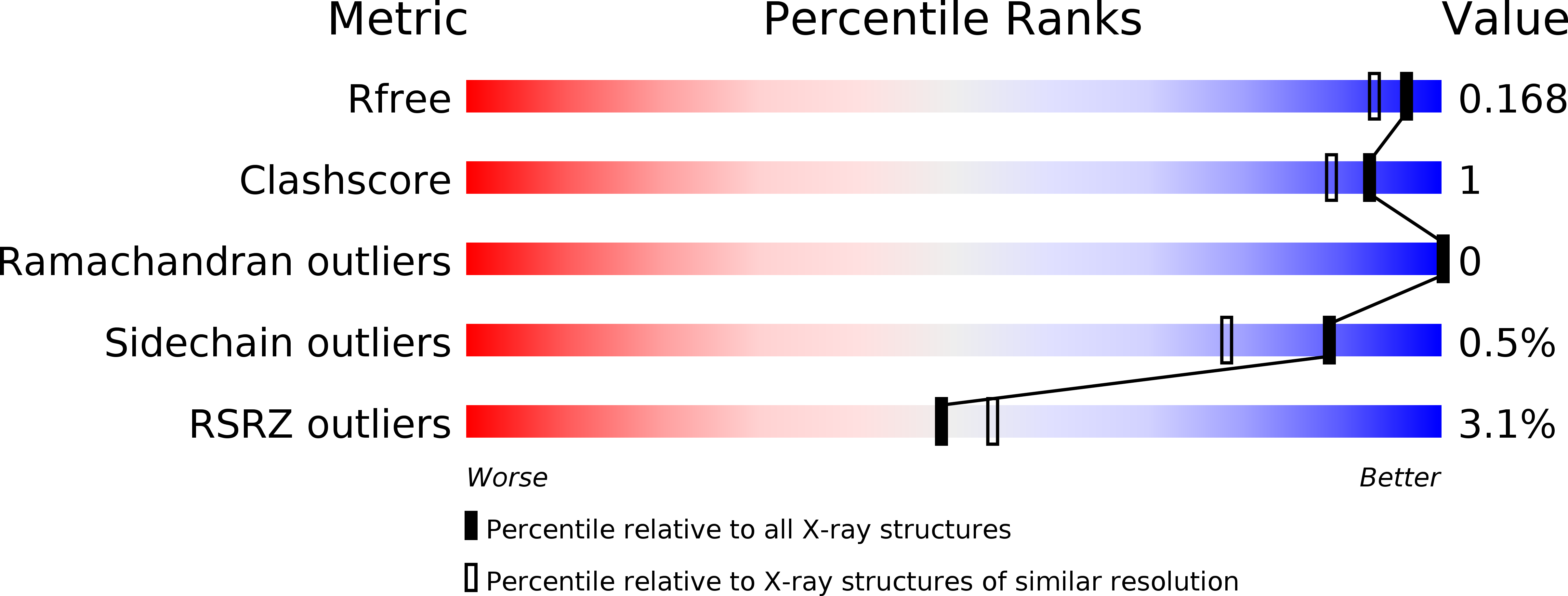
R-Value Free:
0.16
R-Value Work:
0.14
R-Value Observed:
0.14
Space Group:
P 32 2 1