Deposition Date
2014-09-03
Release Date
2015-01-14
Last Version Date
2024-03-20
Entry Detail
Biological Source:
Source Organism(s):
Escherichia coli K-12 (Taxon ID: 83333)
synthetic construct (Taxon ID: 32630)
synthetic construct (Taxon ID: 32630)
Expression System(s):
Method Details:
Experimental Method:
Resolution:
2.30 Å
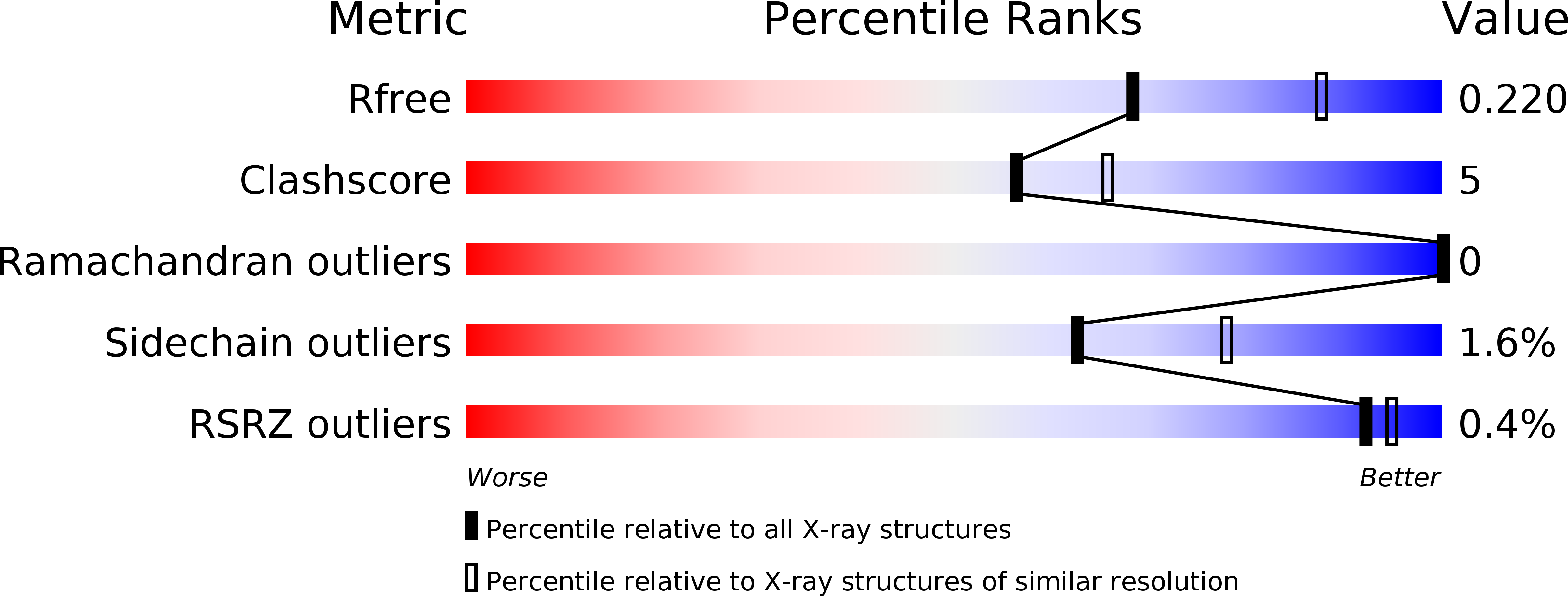
R-Value Free:
0.21
R-Value Work:
0.18
R-Value Observed:
0.18
Space Group:
P 1 21 1