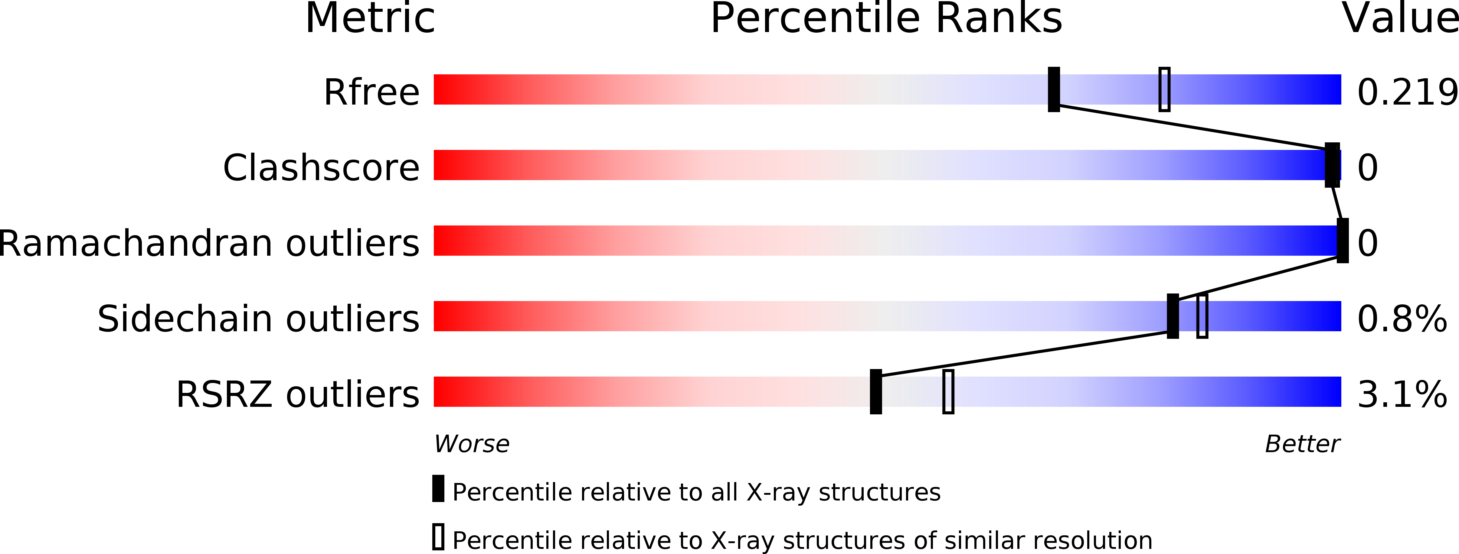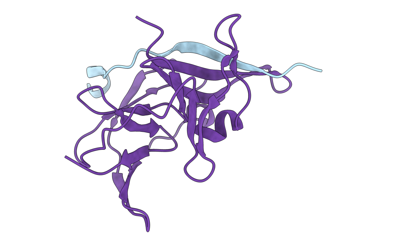
4R8T image
Deposition Date 2014-09-03
Release Date 2014-12-24
Last Version Date 2024-03-20
Entry Detail
PDB ID:
4R8T
Keywords:
Title:
Structure of JEV protease
Biological Source:
Source Organism:
Host Organism:
Method Details:
Experimental Method:
Resolution:
2.13 Å
R-Value Free:
0.21
R-Value Work:
0.16
R-Value Observed:
0.16
Space Group:
P 62
Macromolecular Entities
Polymer Type:polypeptide(L)
Molecule:Serine protease subunit NS2B
Chain IDs:A
Chain Length:21
Number of Molecules:1
Biological Source:Japanese encephalitis virus
Polymer Type:polypeptide(L)
Molecule:NS3
Chain IDs:B
Chain Length:141
Number of Molecules:1
Biological Source:Japanese encephalitis virus
Ligand Molecules
Primary Citation

Abstact

We describe a data collection method that uses a single crystal to solve X-ray structures by native SAD (single-wavelength anomalous diffraction). We solved the structures of 11 real-life examples, including a human membrane protein, a protein-DNA complex and a 266-kDa multiprotein-ligand complex, using this method. The data collection strategy is suitable for routine structure determination and can be implemented at most macromolecular crystallography synchrotron beamlines.

Legend

Protein

Chemical

Disease

Primary Citation of related structures