4R8O image
Entry Detail
PDB ID:
4R8O
Title:
Crystal structure of a DUF3836 family protein (BVU_1206) from Bacteroides vulgatus ATCC 8482 at 2.50 A resolution
Biological Source:
Source Organism:
Host Organism:
PDB Version:
Deposition Date:
2014-09-02
Release Date:
2014-09-17
Method Details:
Experimental Method:
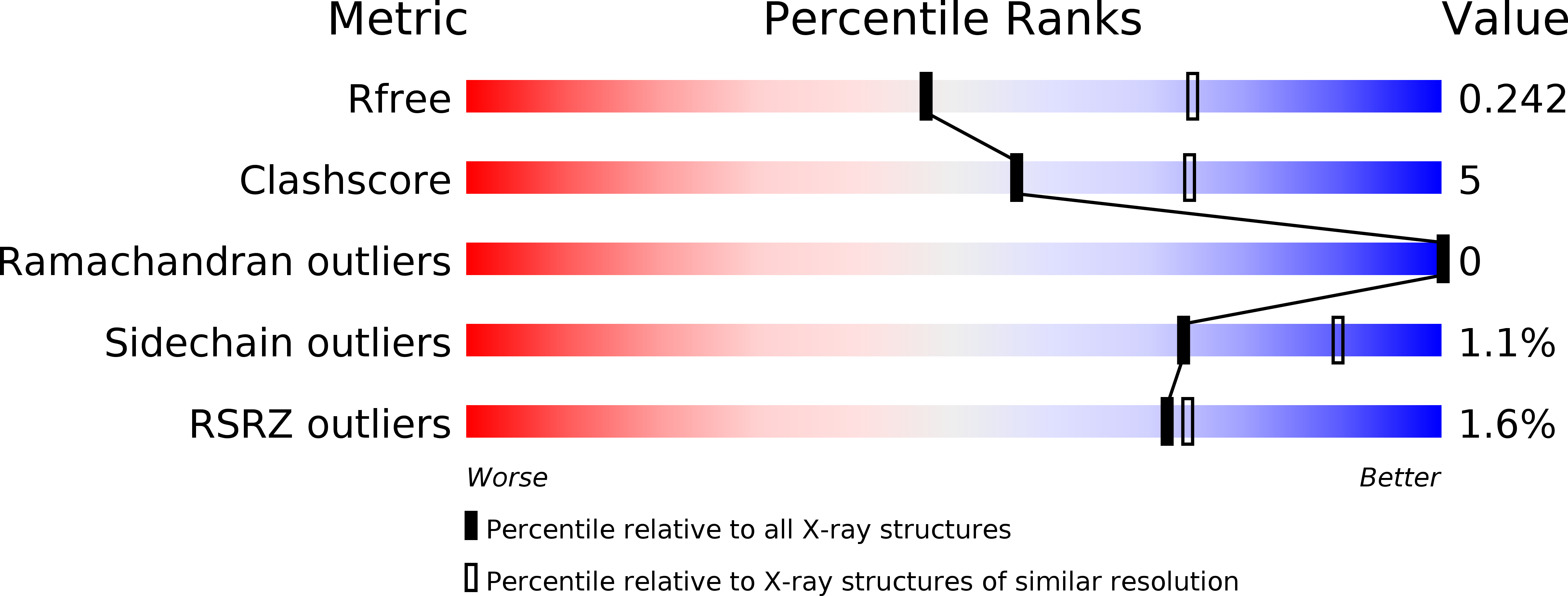
Resolution:
2.50 Å
R-Value Free:
0.24
R-Value Work:
0.17
R-Value Observed:
0.18
Space Group:
P 31 2 1
Macromolecular Entities
Polymer Type:polypeptide(L)
Description:Uncharacterized protein
Chain IDs:A, B
Chain Length:104
Number of Molecules:2
Biological Source:Bacteroides vulgatus
Modified Residue
Compound ID Chain ID Parent Comp ID Details 2D Image
MSE A MET SELENOMETHIONINE
Primary Citation
Structures of single-layer beta-sheet proteins evolved from beta-hairpin repeats.
Protein Sci. 28 1676 1689 (2019)
PMID: 31306512 DOI: 10.1002/pro.3683

Abstact

Free-standing single-layer β-sheets are extremely rare in naturally occurring proteins, even though β-sheet motifs are ubiquitous. Here we report the crystal structures of three homologous, single-layer, anti-parallel β-sheet proteins, comprised of three or four twisted β-hairpin repeats. The structures reveal that, in addition to the hydrogen bond network characteristic of β-sheets, additional hydrophobic interactions mediated by small clusters of residues adjacent to the turns likely play a significant role in the structural stability and compensate for the lack of a compact hydrophobic core. These structures enabled identification of a family of secreted proteins that are broadly distributed in bacteria from the human gut microbiome and are putatively involved in the metabolism of complex carbohydrates. A conserved surface patch, rich in solvent-exposed tyrosine residues, was identified on the concave surface of the β-sheet. These new modular single-layer β-sheet proteins may serve as a new model system for studying folding and design of β-rich proteins.

Legend

Protein

Chemical

Disease

Primary Citation of related structures