4R6C image
Deposition Date 2014-08-24
Release Date 2015-02-04
Last Version Date 2024-10-30
Entry Detail
PDB ID:
4R6C
Keywords:
Title:
X-ray diffraction in temporally and spatially resolved biomolecular science: the X-ray crystal structure of hen egg white lysozyme cocrystallized with Ta6Br12 and then a crystal soaked in K2PtBr6
Biological Source:
Source Organism:
Gallus gallus (Taxon ID: 9031)
Method Details:
Experimental Method:
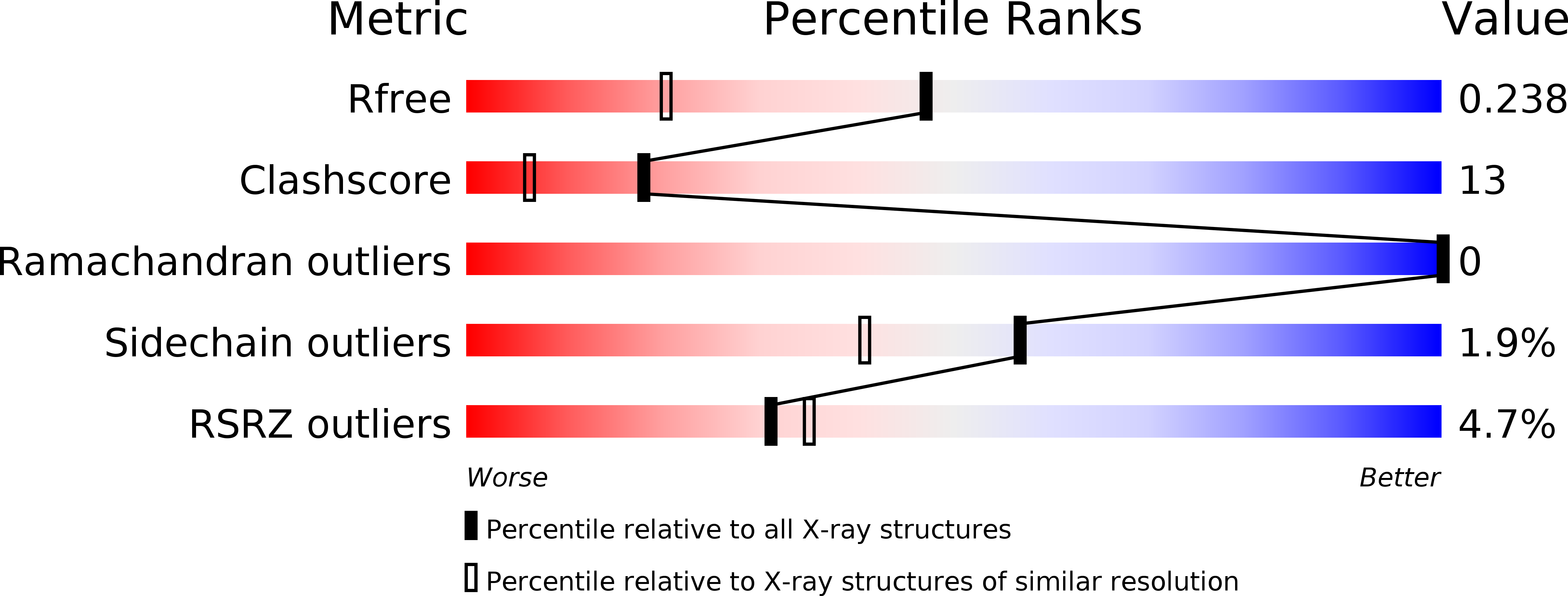
Resolution:
1.70 Å
R-Value Free:
0.23
R-Value Work:
0.18
R-Value Observed:
0.18
Space Group:
P 43 21 2
Macromolecular Entities
Structures with similar UniProt ID
Protein Blast
Polymer Type:polypeptide(L)
Molecule:Lysozyme C
Gene (Uniprot):LYZ
Chain IDs:A
Chain Length:129
Number of Molecules:1
Biological Source:Gallus gallus
Primary Citation
X-ray diffraction in temporally and spatially resolved biomolecular science.
Faraday Discuss 177 429 441 (2015)
PMID: 25605312 DOI: 10.1039/c4fd00166d

Abstact

Time-resolved Laue protein crystallography at the European Synchrotron Radiation Facility (ESRF) opened up the field of sub-nanosecond protein crystal structure analyses. There are a limited number of such time-resolved studies in the literature. Why is this? The X-ray laser now gives us femtosecond (fs) duration pulses, typically 10 fs up to ∼50 fs. Their use is attractive for the fastest time-resolved protein crystallography studies. It has been proposed that single molecules could even be studied with the advantage of being able to measure X-ray diffraction from a 'crystal lattice free' single molecule, with or without temporal resolved structural changes. This is altogether very challenging R&D. So as to assist this effort we have undertaken studies of metal clusters that bind to proteins, both 'fresh' and after repeated X-ray irradiation to assess their X-ray-photo-dynamics, namely Ta6Br12, K2PtI6 and K2PtBr6 bound to a test protein, hen egg white lysozyme. These metal complexes have the major advantage of being very recognisable shapes (pseudo spherical or octahedral) and thereby offer a start to (probably very difficult) single molecule electron density map interpretations, both static and dynamic. A further approach is to investigate the X-ray laser beam diffraction strength of a well scattering nano-cluster; an example from nature being the iron containing ferritin. Electron crystallography and single particle electron microscopy imaging offers alternatives to X-ray structural studies; our structural studies of crustacyanin, a 320 kDa protein carotenoid complex, can be extended either by electron based techniques or with the X-ray laser representing a fascinating range of options. General outlook remarks concerning X-ray, electron and neutron macromolecular crystallography as well as 'NMR crystallography' conclude the article.

Legend

Protein

Chemical

Disease

Primary Citation of related structures