Deposition Date
2014-08-20
Release Date
2014-12-17
Last Version Date
2024-02-28
Entry Detail
PDB ID:
4R4Y
Keywords:
Title:
Structural basis of a point mutation that causes the genetic disease Aspartylglucosaminuria
Biological Source:
Source Organism(s):
Elizabethkingia miricola (Taxon ID: 172045)
Expression System(s):
Method Details:
Experimental Method:
Resolution:
2.10 Å
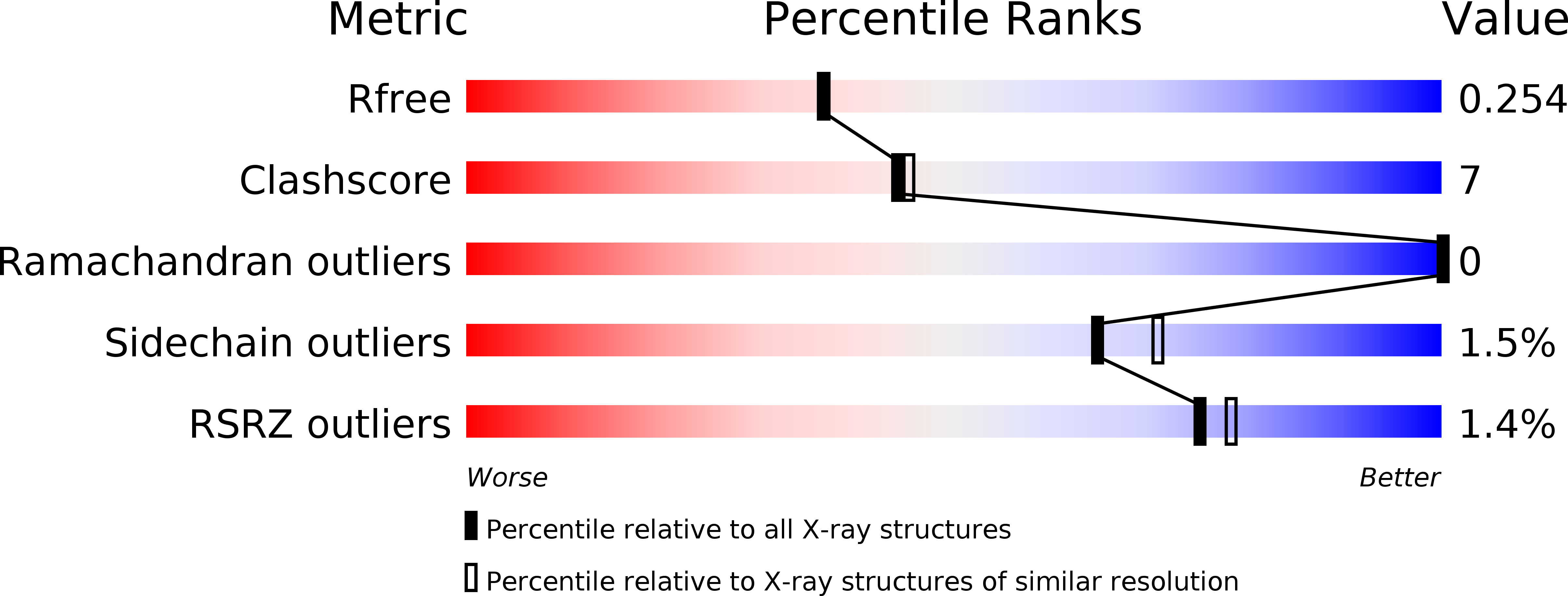
R-Value Free:
0.25
R-Value Work:
0.19
R-Value Observed:
0.19
Space Group:
P 1