4R4P image
Entry Detail
PDB ID:
4R4P
Keywords:
Title:
Crystal Structure of the VS ribozyme-A756G mutant
Biological Source:
Source Organism:
PDB Version:
Deposition Date:
2014-08-19
Release Date:
2015-09-30
Method Details:
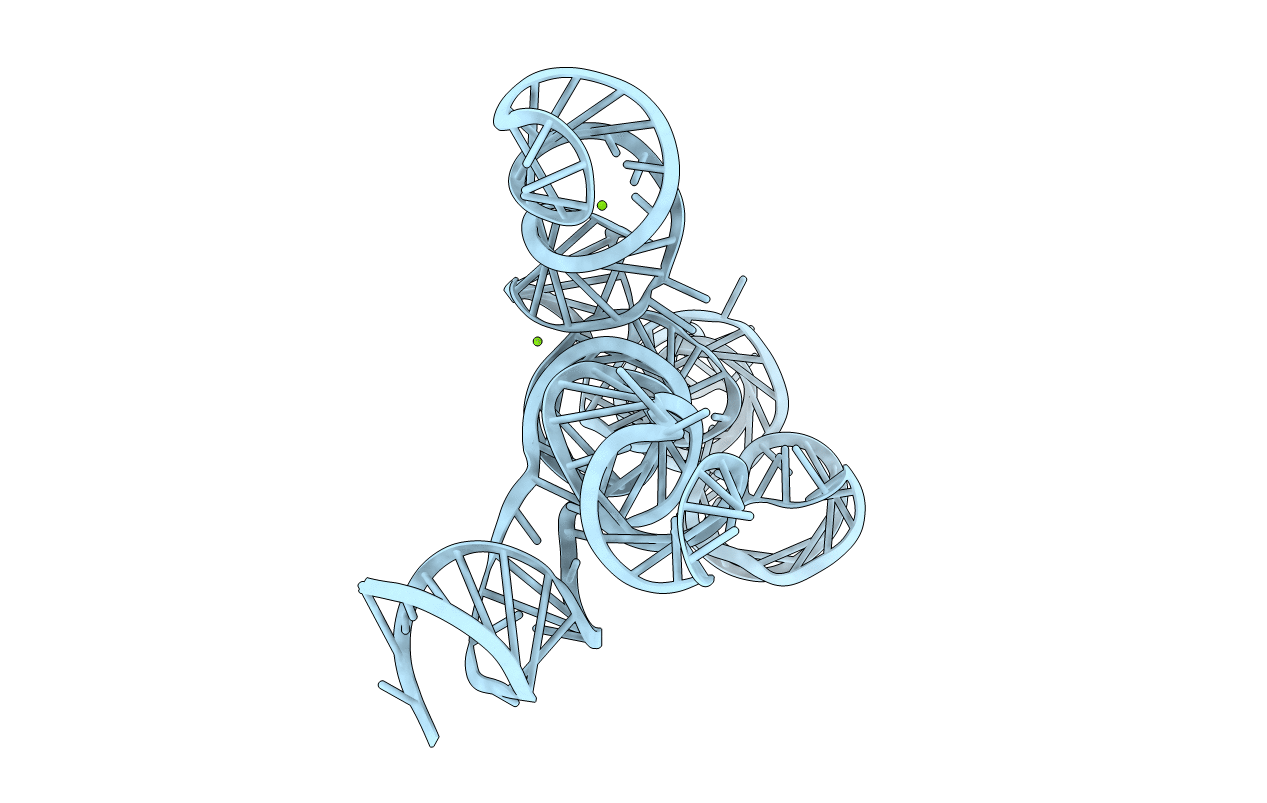
Experimental Method:
Resolution:
3.07 Å
R-Value Free:
0.26
R-Value Work:
0.23
R-Value Observed:
0.23
Space Group:
P 43 21 2
Macromolecular Entities
Polymer Type:polyribonucleotide
Description:VS ribozyme RNA
Mutations:A756G
Chain IDs:A
Chain Length:186
Number of Molecules:1
Biological Source:NEUROSPORA
Modified Residue
Compound ID Chain ID Parent Comp ID Details 2D Image
GDP A G GUANOSINE-5'-DIPHOSPHATE
Ligand Molecules
Primary Citation
Crystal structure of the Varkud satellite ribozyme.
Nat.Chem.Biol. 11 840 846 (2015)
PMID: 26414446 DOI: 10.1038/nchembio.1929

Abstact

The Varkud satellite (VS) ribozyme mediates rolling-circle replication of a plasmid found in the Neurospora mitochondrion. We report crystal structures of this ribozyme from Neurospora intermedia at 3.1 Å resolution, which revealed an intertwined dimer formed by an exchange of substrate helices. In each protomer, an arrangement of three-way helical junctions organizes seven helices into a global fold that creates a docking site for the substrate helix of the other protomer, resulting in the formation of two active sites in trans. This mode of RNA-RNA association resembles the process of domain swapping in proteins and has implications for RNA regulation and evolution. Within each active site, adenine and guanine nucleobases abut the scissile phosphate, poised to serve direct roles in catalysis. Similarities to the active sites of the hairpin and hammerhead ribozymes highlight the functional importance of active-site features, underscore the ability of RNA to access functional architectures from distant regions of sequence space, and suggest convergent evolution.

Legend

Protein

Chemical

Disease

Primary Citation of related structures