Deposition Date
2014-08-19
Release Date
2015-09-23
Last Version Date
2024-04-03
Entry Detail
PDB ID:
4R4M
Keywords:
Title:
Crystal structure of C42L cGMP dependent protein kinase I alpha (PKGI alpha) leucine zipper
Biological Source:
Source Organism(s):
Homo sapiens (Taxon ID: 9606)
Expression System(s):
Method Details:
Experimental Method:
Resolution:
1.92 Å
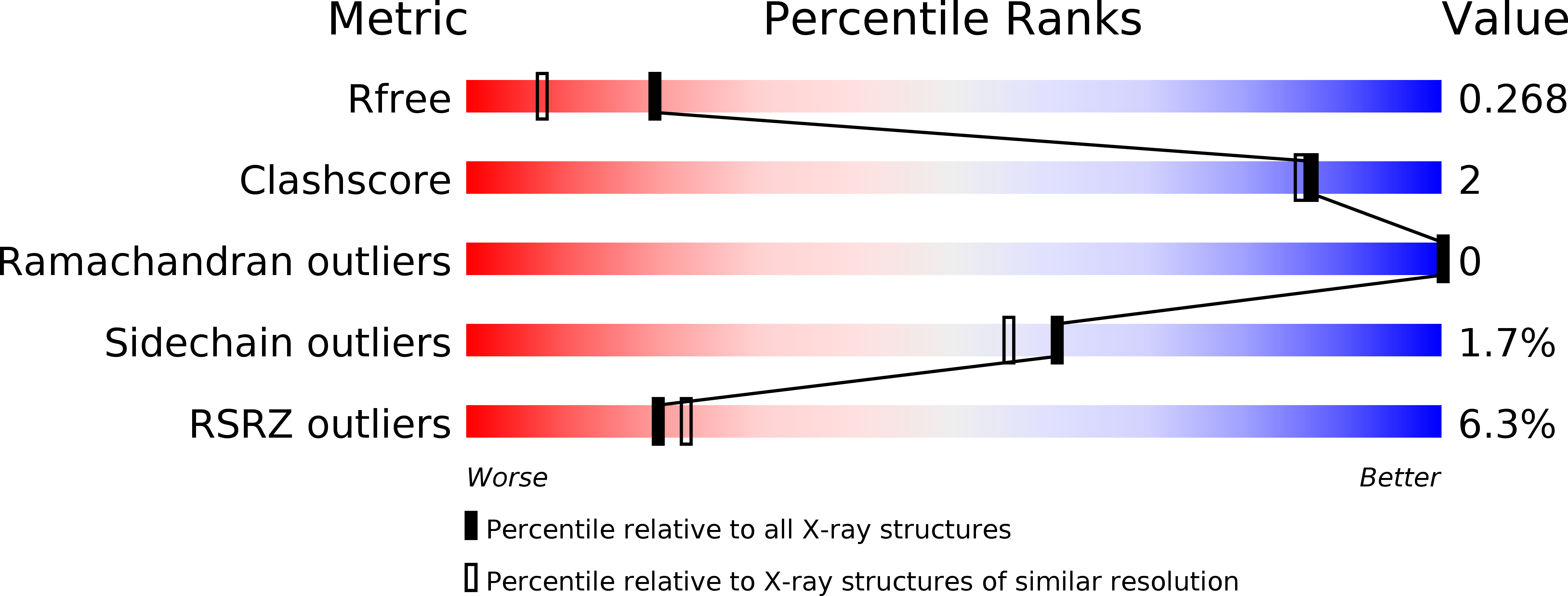
R-Value Free:
0.26
R-Value Work:
0.22
R-Value Observed:
0.22
Space Group:
P 62 2 2