
Deposition Date
2014-08-19
Release Date
2015-08-19
Last Version Date
2024-03-20
Entry Detail
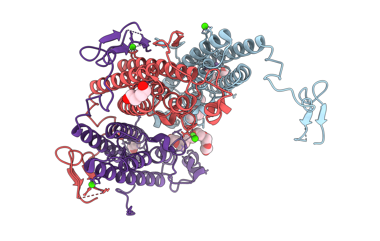
PDB ID:
4R42
Keywords:
Title:
Crystal structure of KatB, a manganese catalase from Anabaena PCC7120
Biological Source:
Source Organism(s):
Nostoc sp. PCC 7120 (Taxon ID: 103690)
Expression System(s):
Method Details:
Experimental Method:
Resolution:
1.90 Å
R-Value Free:
0.15
R-Value Work:
0.13
R-Value Observed:
0.13
Space Group:
P 41 21 2
