4R30 image
Deposition Date 2014-08-13
Release Date 2014-12-31
Last Version Date 2023-09-20
Entry Detail
PDB ID:
4R30
Keywords:
Title:
Structure of human laforin dual specificity phosphatase domain
Biological Source:
Source Organism:
Homo sapiens (Taxon ID: 9606)
Host Organism:
Method Details:
Experimental Method:
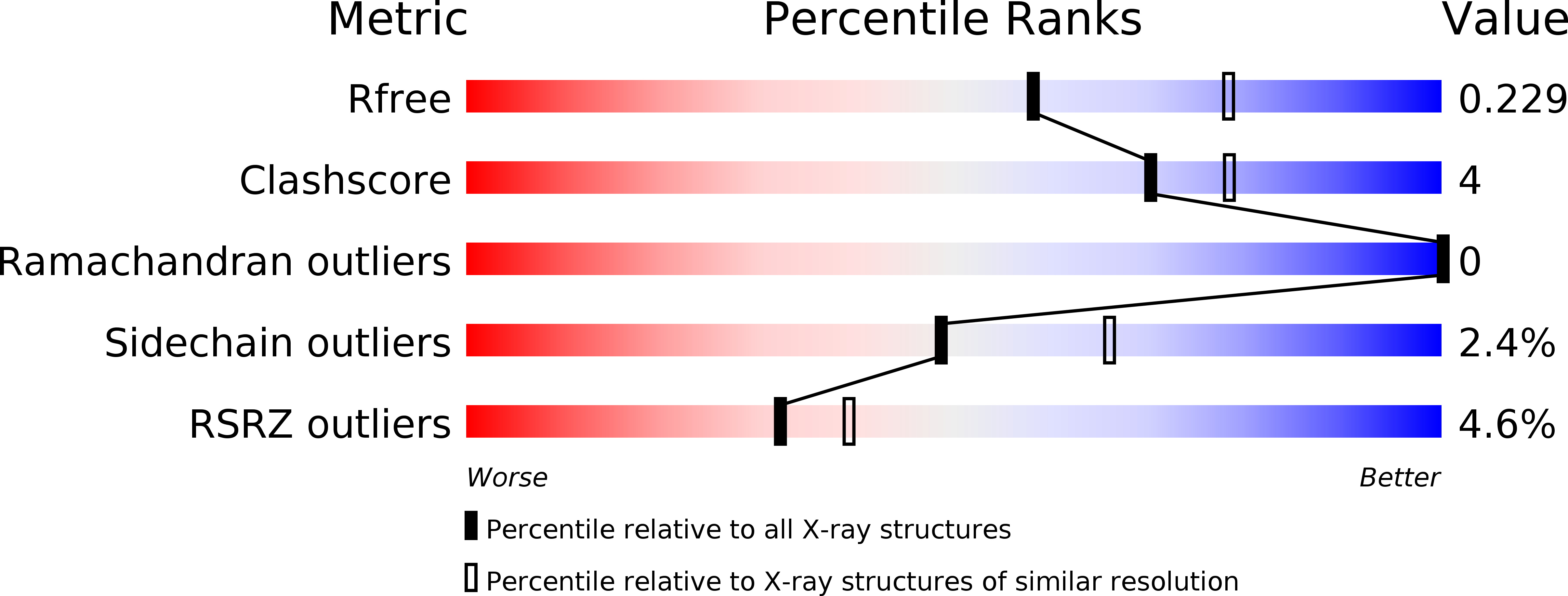
Resolution:
2.30 Å
R-Value Free:
0.21
R-Value Work:
0.18
R-Value Observed:
0.18
Space Group:
I 41
Macromolecular Entities
Structures with similar UniProt ID
Protein Blast
Polymer Type:polypeptide(L)
Molecule:Laforin
Gene (Uniprot):EPM2A
Mutagens:C266S
Chain IDs:A, B, C, D
Chain Length:184
Number of Molecules:4
Biological Source:Homo sapiens
Primary Citation
Dimeric quaternary structure of human laforin.
J.Biol.Chem. 290 4552 4559 (2015)
PMID: 25538239 DOI: 10.1074/jbc.M114.627406

Abstact

The phosphatase laforin removes phosphate groups from glycogen during biosynthetic activity. Loss-of-function mutations in the gene encoding laforin is the predominant cause of Lafora disease, a fatal form of progressive myoclonic epilepsy. Here, we used hybrid structural methods to determine the molecular architecture of human laforin. We found that laforin adopts a dimeric quaternary structure, topologically similar to the prototypical dual specificity phosphatase VH1. The interface between the laforin carbohydrate-binding module and the dual specificity phosphatase domain generates an intimate substrate-binding crevice that allows for recognition and dephosphorylation of phosphomonoesters of glucose. We identify novel molecular determinants in the laforin active site that help decipher the mechanism of glucan phosphatase activity.

Legend

Protein

Chemical

Disease

Primary Citation of related structures