Deposition Date
2014-08-13
Release Date
2014-12-10
Last Version Date
2023-09-20
Entry Detail
PDB ID:
4R2X
Keywords:
Title:
Unique conformation of uridine and asymmetry of the hexameric molecule revealed in the high-resolution structures of Shewanella oneidensis uridine phosphorylase in the free form and in complex with uridine
Biological Source:
Source Organism(s):
Shewanella oneidensis (Taxon ID: 211586)
Expression System(s):
Method Details:
Experimental Method:
Resolution:
0.93 Å
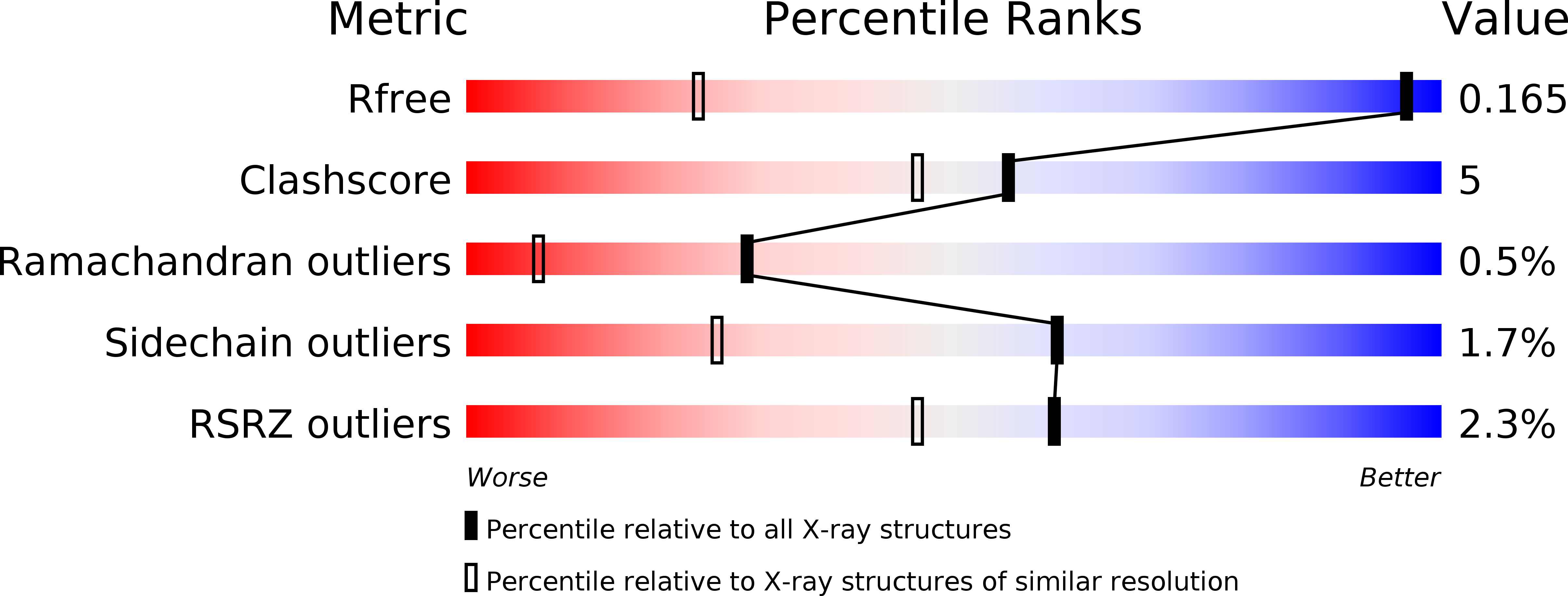
R-Value Free:
0.16
R-Value Work:
0.14
R-Value Observed:
0.15
Space Group:
P 1 21 1