4R19 image
Entry Detail
PDB ID:
4R19
Keywords:
Title:
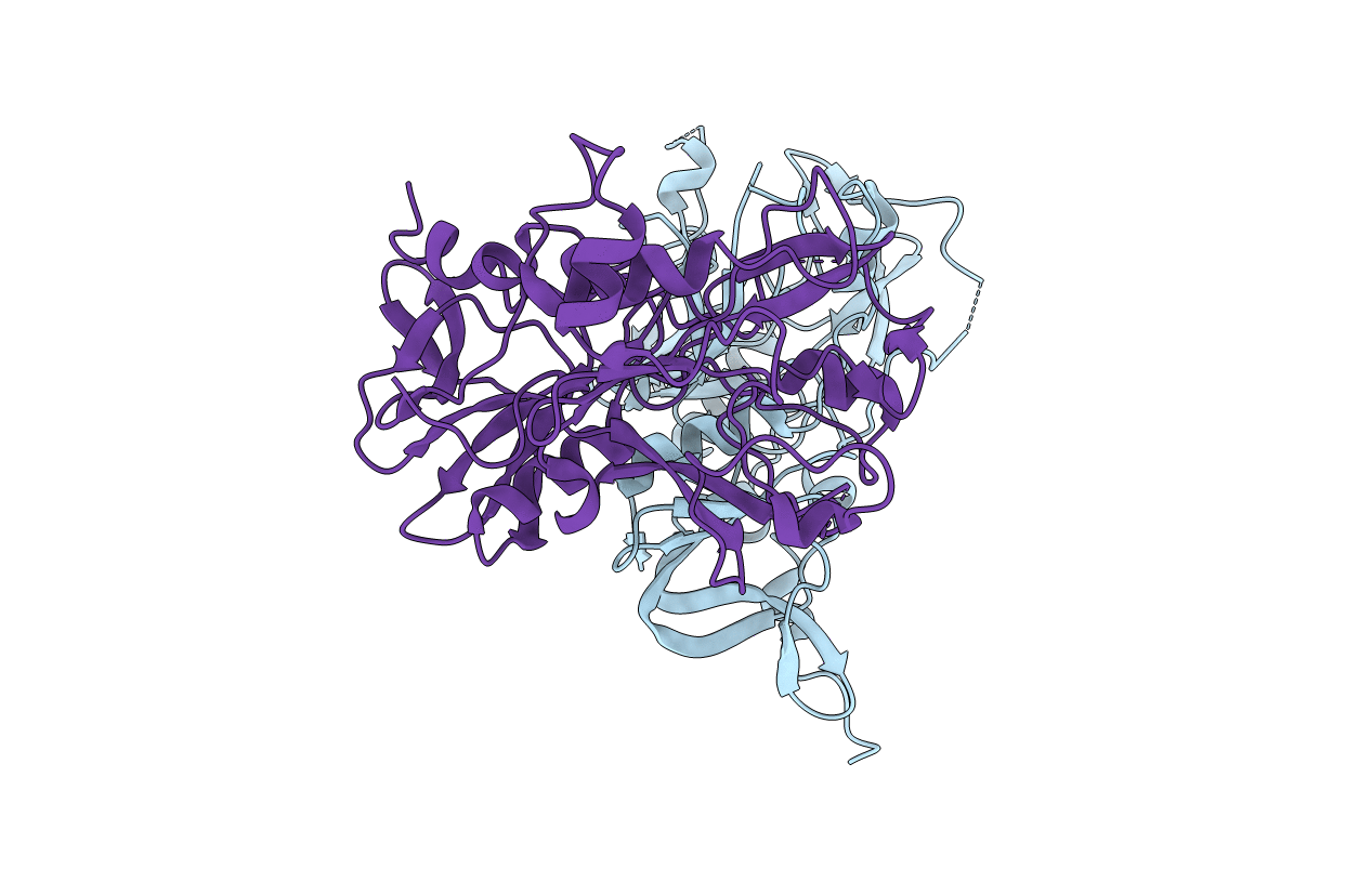
Crystal Structure of 3D7 strain Plasmodium falciparum AMA1
Biological Source:
Source Organism:
Host Organism:
PDB Version:
Deposition Date:
2014-08-04
Release Date:
2015-01-14
Method Details:
Experimental Method:
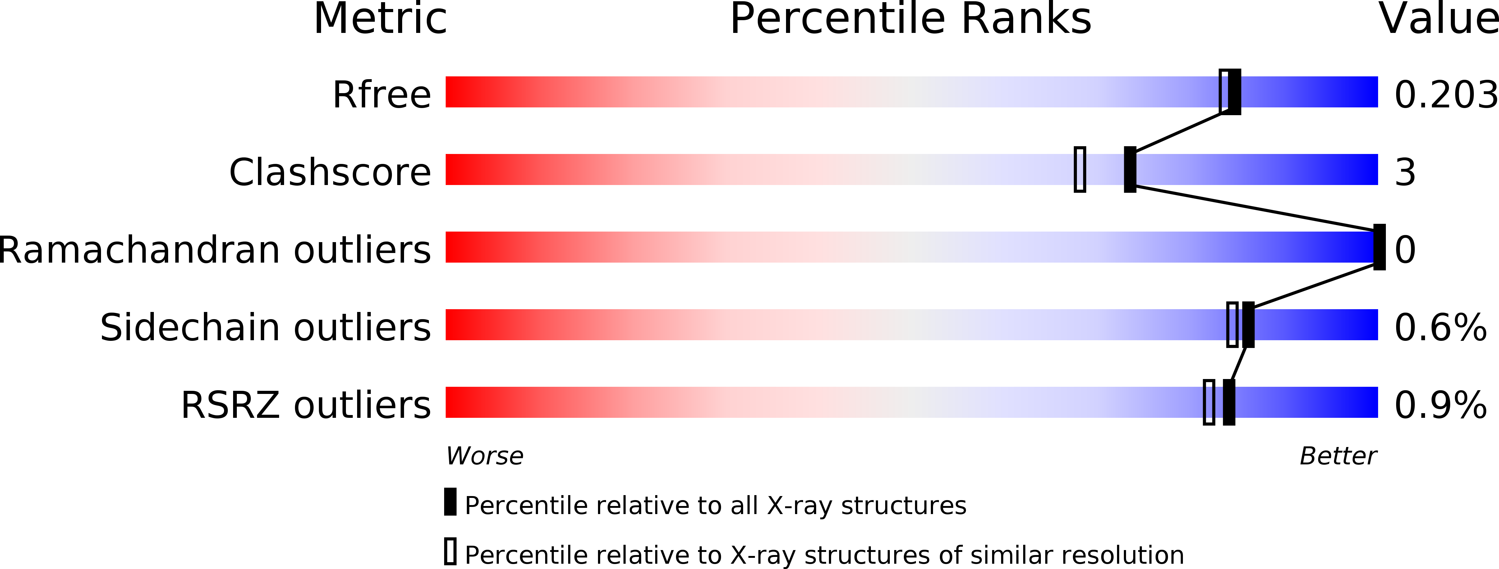
Resolution:
1.80 Å
R-Value Free:
0.20
R-Value Work:
0.17
R-Value Observed:
0.17
Space Group:
P 31
Macromolecular Entities
Polymer Type:polypeptide(L)
Description:Apical membrane antigen 1, AMA1
Chain IDs:A, B
Chain Length:335
Number of Molecules:2
Biological Source:Plasmodium falciparum 3D7
Primary Citation
Structure and Dynamics of Apical Membrane Antigen 1 from Plasmodium falciparum FVO.
Biochemistry 53 7310 7320 (2014)
PMID: 25360546 DOI: 10.1021/bi5012089

Abstact

Apical membrane antigen 1 (AMA1) interacts with RON2 to form a protein complex that plays a key role in the invasion of host cells by malaria parasites. Blocking this protein-protein interaction represents a potential route to controlling malaria and related parasitic diseases, but the polymorphic nature of AMA1 has proven to be a major challenge to vaccine-induced antibodies and peptide inhibitors exerting strain-transcending inhibitory effects. Here we present the X-ray crystal structure of AMA1 domains I and II from Plasmodium falciparum strain FVO. We compare our new structure to those of AMA1 from P. falciparum 3D7 and Plasmodium vivax. A combination of normalized B factor analysis and computational methods has been used to investigate the flexibility of the domain I loops and how this correlates with their roles in determining the strain specificity of human antibody responses and inhibitory peptides. We also investigated the domain II loop, a key region involved in inhibitor binding, by comparison of multiple AMA1 crystal structures. Collectively, these results provide valuable insights that should contribute to the design of strain-transcending agents targeting P. falciparum AMA1.

Legend

Protein

Chemical

Disease

Primary Citation of related structures