4QYP image
Deposition Date 2014-07-25
Release Date 2014-12-10
Last Version Date 2023-09-20
Entry Detail
PDB ID:
4QYP
Title:
The Crystal Structures of holo-wt human Cellular Retinol Binding protein II (hCRBPII) bound to Retinal
Biological Source:
Source Organism:
Homo sapiens (Taxon ID: 9606)
Host Organism:
Method Details:
Experimental Method:
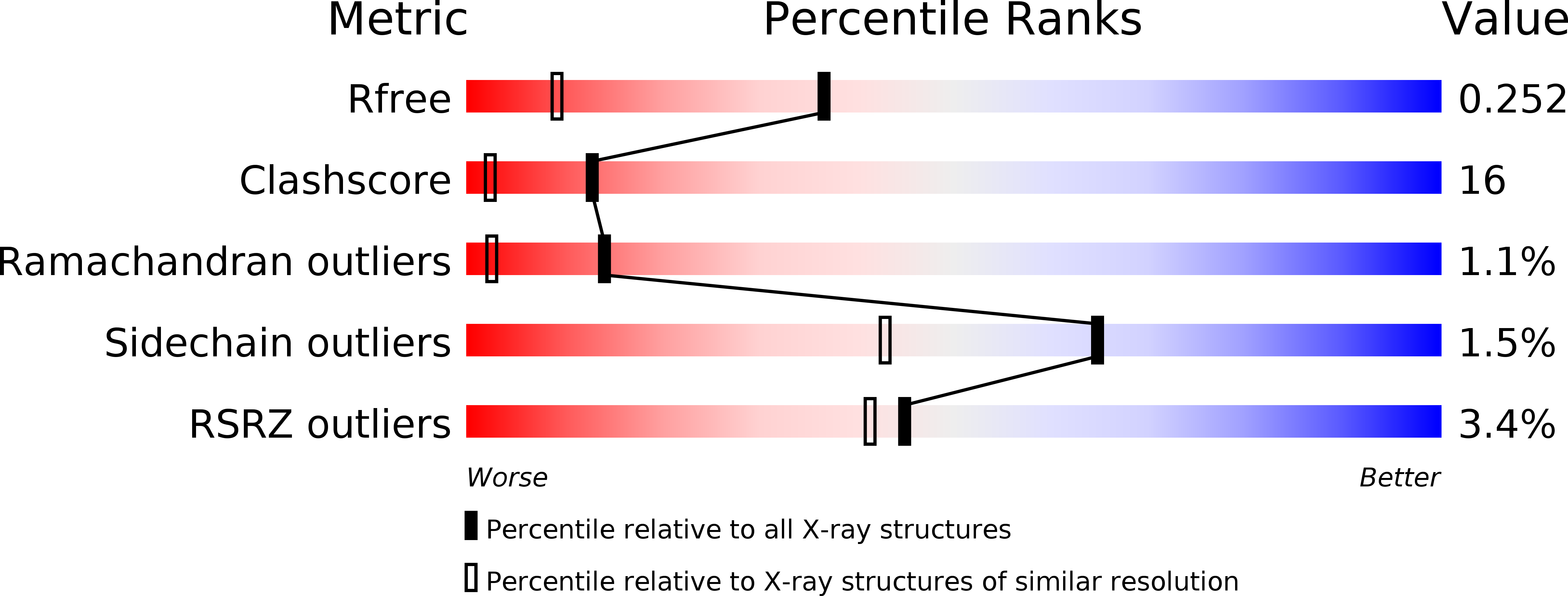
Resolution:
1.62 Å
R-Value Free:
0.25
R-Value Work:
0.20
R-Value Observed:
0.20
Space Group:
P 1
Macromolecular Entities
Polymer Type:polypeptide(L)
Molecule:Retinol-binding protein 2
Gene (Uniprot):RBP2
Chain IDs:A, B, C, D
Chain Length:133
Number of Molecules:4
Biological Source:Homo sapiens
Primary Citation
Structures of holo wild-type human cellular retinol-binding protein II (hCRBPII) bound to retinol and retinal.
Acta Crystallogr.,Sect.D 70 3226 3232 (2014)
PMID: 25478840 DOI: 10.1107/S1399004714023839

Abstact

Cellular retinol-binding proteins (CRBPs) I and II, which are members of the intracellular lipid-binding protein (iLBP) family, are retinoid chaperones that are responsible for the intracellular transport and delivery of both retinol and retinal. Although structures of retinol-bound CRBPI and CRBPII are known, no structure of a retinal-bound CRBP has been reported. In addition, the retinol-bound human CRBPII (hCRBPII) structure shows partial occupancy of a noncanonical conformation of retinol in the binding pocket. Here, the structure of retinal-bound hCRBPII and the structure of retinol-bound hCRBPII with retinol fully occupying the binding pocket are reported. It is further shown that the retinoid derivative seen in both the zebrafish CRBP and the hCRBPII structures is likely to be the product of flux-dependent and wavelength-dependent X-ray damage during data collection. The structures of retinoid-bound CRBPs are compared and contrasted, and rationales for the differences in binding affinities for retinal and retinol are provided.

Legend

Protein

Chemical

Disease

Primary Citation of related structures