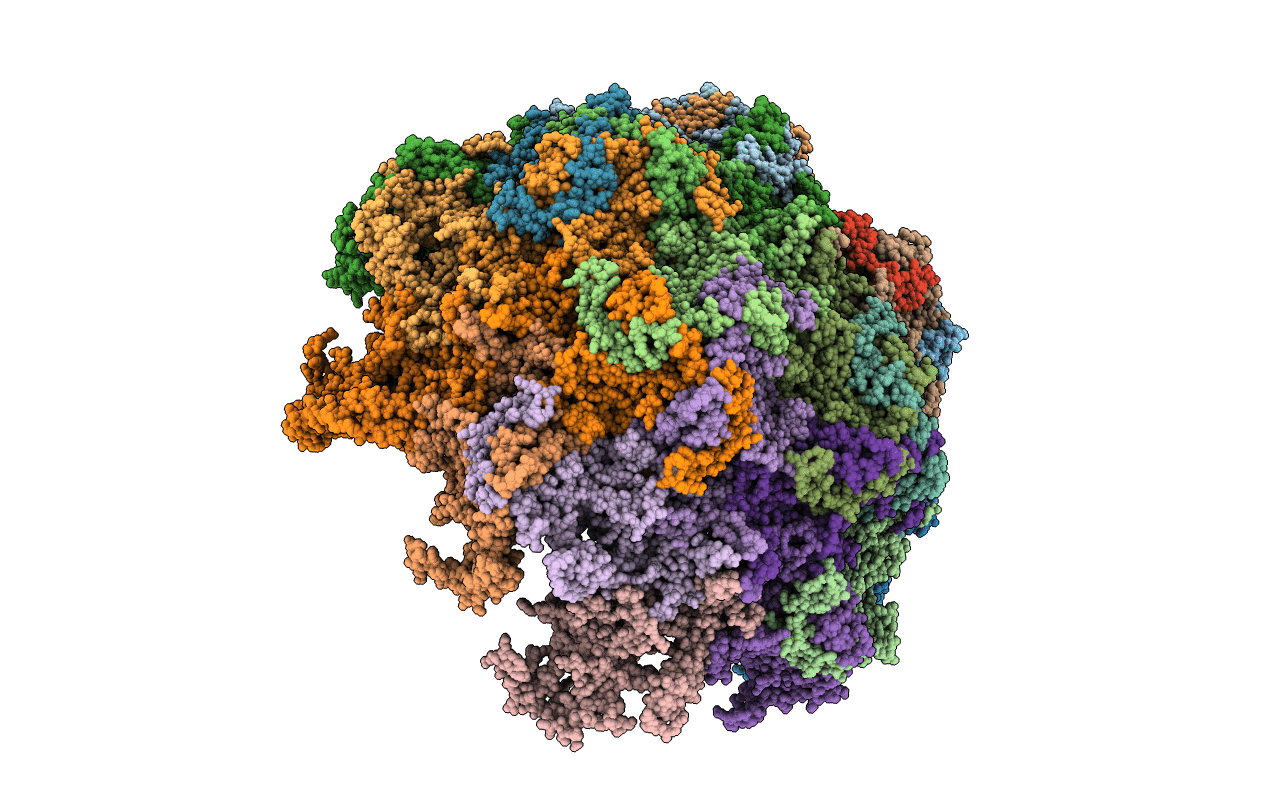
4QYK image
Entry Detail
PDB ID:
4QYK
Keywords:
Title:
Crystal structure of a canine parvovirus variant
Biological Source:
Host Organism:
PDB Version:
Deposition Date:
2014-07-24
Release Date:
2014-12-10
Method Details:
Experimental Method:
Resolution:
3.50 Å
R-Value Free:
0.22
R-Value Work:
0.18
R-Value Observed:
0.18
Space Group:
P 42 21 2
Macromolecular Entities
Polymer Type:polypeptide(L)
Description:Capsid protein VP1
Chain IDs:A, B (auth: E), C (auth: J), D (auth: N), E (auth: Q), F (auth: B), G (auth: C), H (auth: D), I (auth: F), J (auth: G), K (auth: H), L (auth: I), M (auth: K), N (auth: L), O (auth: M), P (auth: O), Q (auth: P), R, S, T, U, V, W, X, Y, Z, AA (auth: a), BA (auth: b), CA (auth: c), DA (auth: d)
Chain Length:584
Number of Molecules:30
Biological Source:Feline panleukopenia virus
Ligand Molecules
Primary Citation
Global Displacement of Canine Parvovirus by a Host-Adapted Variant: Structural Comparison between Pandemic Viruses with Distinct Host Ranges.
J.Virol. 89 1909 1912 (2015)
PMID: 25410876 DOI: 10.1128/JVI.02611-14

Abstact

Canine parvovirus type 2 (CPV-2) emerged in 1978 and spread worldwide within 2 years. Subsequently, CPV-2 was completely replaced by the variant CPV-2a, which is characterized by four specific capsid (VP2) mutations. The X-ray crystal structure of the CPV-2a capsid shows that each mutation confers small local changes. The loss of a hydrogen bond and introduction of a glycine residue likely introduce flexibility to sites that control interactions with the host receptor, antibodies, and sialic acids.

Legend

Protein

Chemical

Disease

Primary Citation of related structures