
Deposition Date
2014-07-11
Release Date
2014-12-10
Last Version Date
2023-11-08
Entry Detail
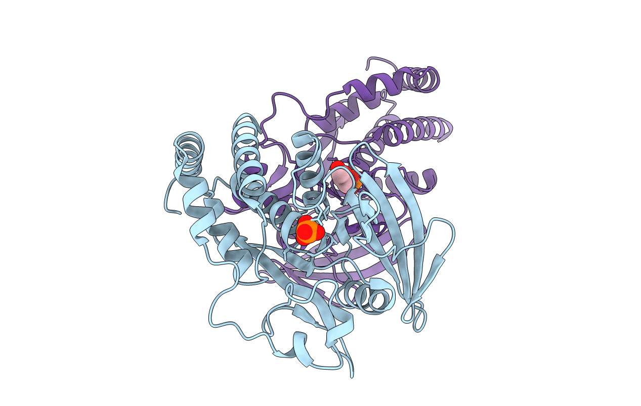
PDB ID:
4QUN
Keywords:
Title:
Crystal structure of the PTPN3 (PTPH1) catalytic domain C842S mutant
Biological Source:
Source Organism(s):
Homo sapiens (Taxon ID: 9606)
Expression System(s):
Method Details:
Experimental Method:
Resolution:
1.86 Å
R-Value Free:
0.20
R-Value Work:
0.17
R-Value Observed:
0.17
Space Group:
C 2 2 21
