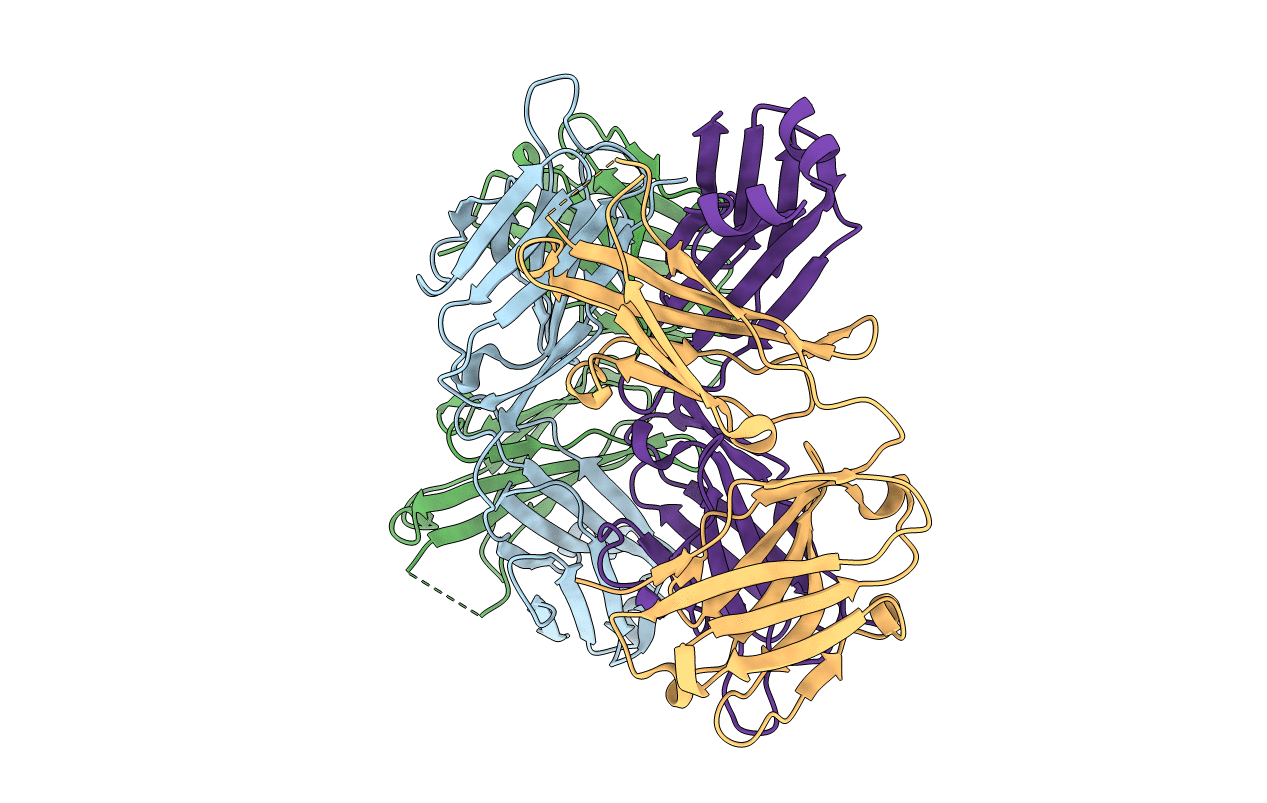
4QT5 image
Entry Detail
PDB ID:
4QT5
Keywords:
Title:
Crystal Structure of 3BD10: A Monoclonal Antibody against the TSH Receptor
Biological Source:
Source Organism:
Host Organism:
PDB Version:
Deposition Date:
2014-07-07
Release Date:
2015-04-01
Method Details:
Experimental Method:
Resolution:
2.50 Å
R-Value Free:
0.27
R-Value Work:
0.23
R-Value Observed:
0.23
Space Group:
P 21 21 21
Macromolecular Entities
Polymer Type:polypeptide(L)
Description:3BD10 mouse monoclonal antibody, heavy chain
Chain IDs:B (auth: H), D (auth: B)
Chain Length:213
Number of Molecules:2
Biological Source:Mus musculus
Polymer Type:polypeptide(L)
Description:3BD10 mouse monoclonal antibody, light chain
Chain IDs:A (auth: L), C (auth: A)
Chain Length:216
Number of Molecules:2
Biological Source:Mus musculus
Primary Citation
Crystal structure of a TSH receptor monoclonal antibody: insight into Graves' disease pathogenesis.
Mol.Endocrinol. 29 99 107 (2015)
PMID: 25419797 DOI: 10.1210/me.2014-1257

Abstact

The TSH receptor (TSHR) A-subunit is more effective than the holoreceptor in inducing thyroid-stimulating antibodies (TSAb) that cause Graves' disease. A puzzling phenomenon is that 2 recombinant, eukaryotic forms of A-subunits (residues 22-289), termed active and inactive, are recognized mutually exclusively by pathogenic TSAb and mouse monoclonal antibody 3BD10, respectively. Understanding the structural difference between these TSHR A-subunit forms could provide insight into Graves' disease pathogenesis. The 3-dimensional structure of the active A-subunit (in complex with a human TSAb Fab, M22) is known, but the structural difference with inactive A-subunits is unknown. We solved the 3BD10 Fab 3-dimensional crystal structure. Guided by prior knowledge of a portion of its epitope, 3BD10 docked in silico with the known active TSHR-289 monomeric structure. Because both TSAb and 3BD10 recognize the active TSHR A-subunit monomer, this form of the molecule can be excluded as the basis for the active-inactive dichotomy, suggesting, instead a role for A-subunit quaternary structure. Indeed, in silico analysis revealed that M22, but not 3BD10, bound to a TSHR-289 trimer. In contrast, 3BD10, but not M22, bound to a TSHR-289 dimer. The validity of these models is supported experimentally by the temperature-dependent balance between active and inactive TSHR-289. In summary, we provide evidence for a structural basis to explain the conformational heterogeneity of TSHR A-subunits (TSHR-289). The pathophysiologic importance of these findings is that affinity maturation of pathogenic TSAb in Graves' disease is likely to involve a trimer of the shed TSHR A-subunit.

Legend

Protein

Chemical

Disease

Primary Citation of related structures