4QT4 image
Deposition Date 2014-07-07
Release Date 2014-08-06
Last Version Date 2023-11-08
Entry Detail
PDB ID:
4QT4
Keywords:
Title:
Crystal structure of Peptidyl-tRNA hydrolase from a Gram-positive bacterium, Streptococcus pyogenes at 2.19 Angstrom resolution shows the Closed Structure of the Substrate Binding Cleft
Biological Source:
Source Organism:
Host Organism:
Method Details:
Experimental Method:
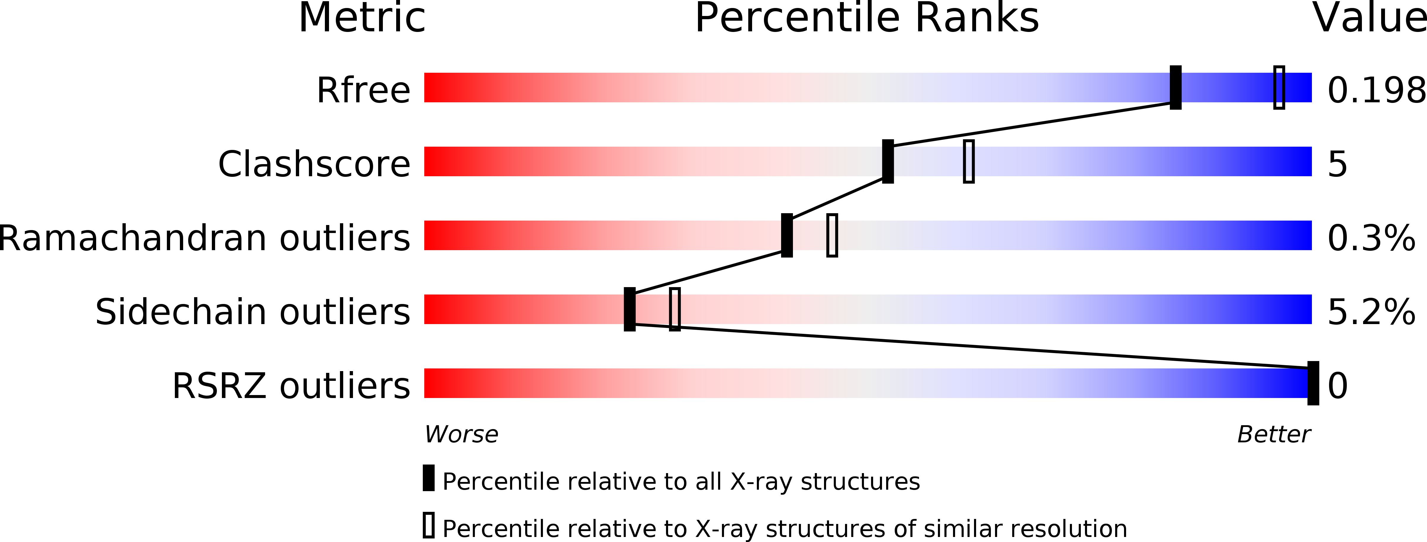
Resolution:
2.19 Å
R-Value Free:
0.19
R-Value Work:
0.16
R-Value Observed:
0.17
Space Group:
P 1
Macromolecular Entities
Polymer Type:polypeptide(L)
Molecule:Peptidyl-tRNA hydrolase
Gene (Uniprot):pth
Chain IDs:A, B
Chain Length:189
Number of Molecules:2
Biological Source:Streptococcus pyogenes NZ131
Primary Citation
Crystal structure of peptidyl-tRNA hydrolase from a Gram-positive bacterium, Streptococcus pyogenes at 2.19 angstrom resolution shows the closed structure of the substrate-binding cleft.
FEBS Open Bio 4 915 922 (2014)
PMID: 25389518 DOI: 10.1016/j.fob.2014.10.010

Abstact

Peptidyl-tRNA hydrolase (Pth) catalyses the release of tRNA and peptide components from peptidyl-tRNA molecules. Pth from a Gram-positive bacterium Streptococcus pyogenes (SpPth) was cloned, expressed, purified and crystallised. Three-dimensional structure of SpPth was determined by X-ray crystallography at 2.19 Å resolution. Structure determination showed that the asymmetric unit of the unit cell contained two crystallographically independent molecules, designated A and B. The superimposition of C(α) traces of molecules A and B showed an r.m.s. shift of 0.4 Å, indicating that the structures of two crystallographically independent molecules were identical. The polypeptide chain of SpPth adopted an overall α/β conformation. The substrate-binding cleft in SpPth is formed with three loops: the gate loop, Ile91-Leu102; the base loop, Gly108-Gly115; and the lid loop, Gly136-Gly150. Unlike in the structures of Pth from Gram-negative bacteria, the entry to the cleft in the structure of SpPth appeared to be virtually closed. However, the conformations of the active site residues were found to be similar.

Legend

Protein

Chemical

Disease

Primary Citation of related structures