4QSG image
Deposition Date 2014-07-03
Release Date 2014-11-12
Last Version Date 2024-03-20
Entry Detail
PDB ID:
4QSG
Title:
Crystal structure of gas vesicle protein GvpF from Microcystis aeruginosa
Biological Source:
Source Organism:
Host Organism:
Method Details:
Experimental Method:
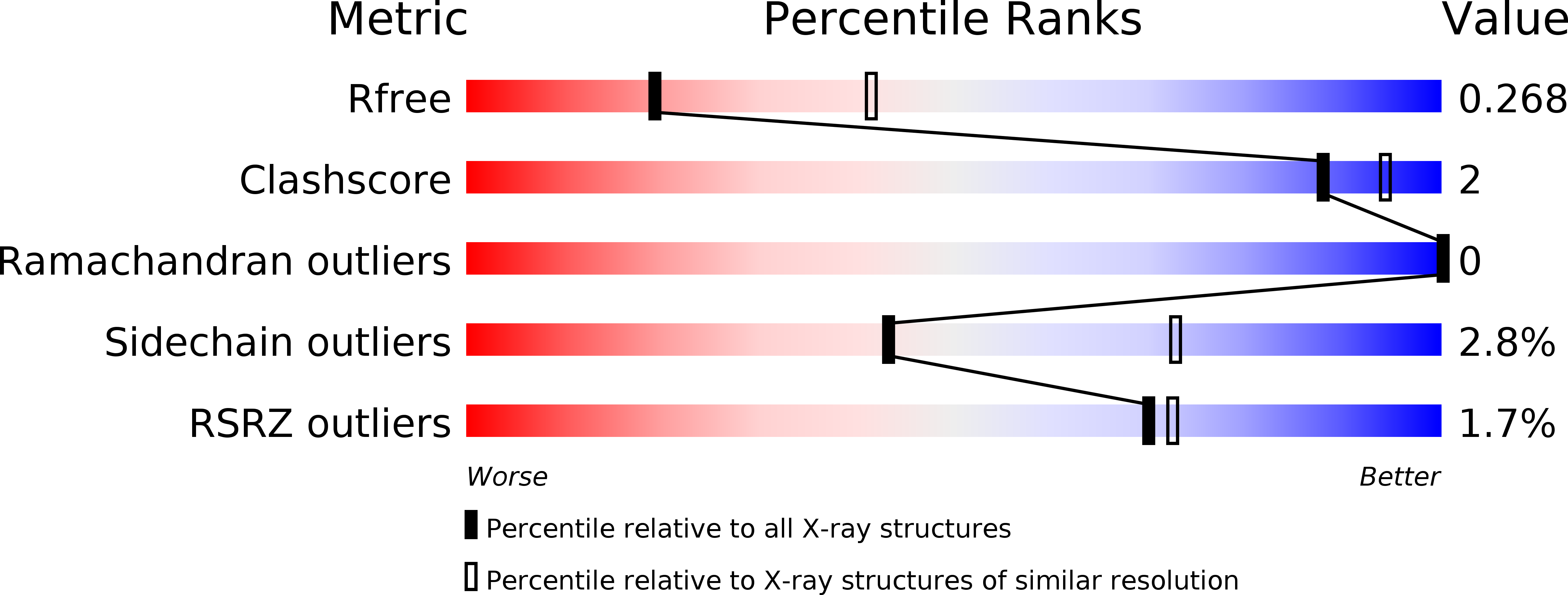
Resolution:
2.70 Å
R-Value Free:
0.27
R-Value Work:
0.22
R-Value Observed:
0.23
Space Group:
P 32 2 1
Macromolecular Entities
Polymer Type:polypeptide(L)
Molecule:Gas vesicle protein
Gene (Uniprot):gvpF
Chain IDs:A
Chain Length:253
Number of Molecules:1
Biological Source:Microcystis aeruginosa PCC 7806
Primary Citation
Structure of the gas vesicle protein GvpF from the cyanobacterium Microcystis aeruginosa.
Acta Crystallogr.,Sect.D 70 3013 3022 (2014)
PMID: 25372690 DOI: 10.1107/S1399004714021312

Abstact

Gas vesicles are gas-filled proteinaceous organelles that provide buoyancy for bacteria and archaea. A gene cluster that is highly conserved in various species encodes about 8-14 proteins (Gvp proteins) that are involved in the formation of gas vesicles. Here, the first crystal structure of the gas vesicle protein GvpF from Microcystis aeruginosa PCC 7806 is reported at 2.7 Å resolution. GvpF is composed of two structurally distinct domains (the N-domain and C-domain), both of which display an α+β class overall structure. The N-domain adopts a novel fold, whereas the C-domain has a modified ferredoxin fold with an apparent variation owing to an extension region consisting of three sequential helices. The two domains pack against each other via interactions with a C-terminal tail that is conserved among cyanobacteria. Taken together, it is concluded that the overall architecture of GvpF presents a novel fold. Moreover, it is shown that GvpF is most likely to be a structural protein that is localized at the gas-facing surface of the gas vesicle by immunoblotting and immunogold labelling-based tomography.

Legend

Protein

Chemical

Disease

Primary Citation of related structures