Deposition Date
2014-07-01
Release Date
2015-06-10
Last Version Date
2024-02-28
Entry Detail
PDB ID:
4QRM
Keywords:
Title:
crystal structure of a binary complex of FliM-FliG middle domains from T.maritima
Biological Source:
Source Organism(s):
Thermotoga maritima (Taxon ID: 243274)
Expression System(s):
Method Details:
Experimental Method:
Resolution:
4.32 Å
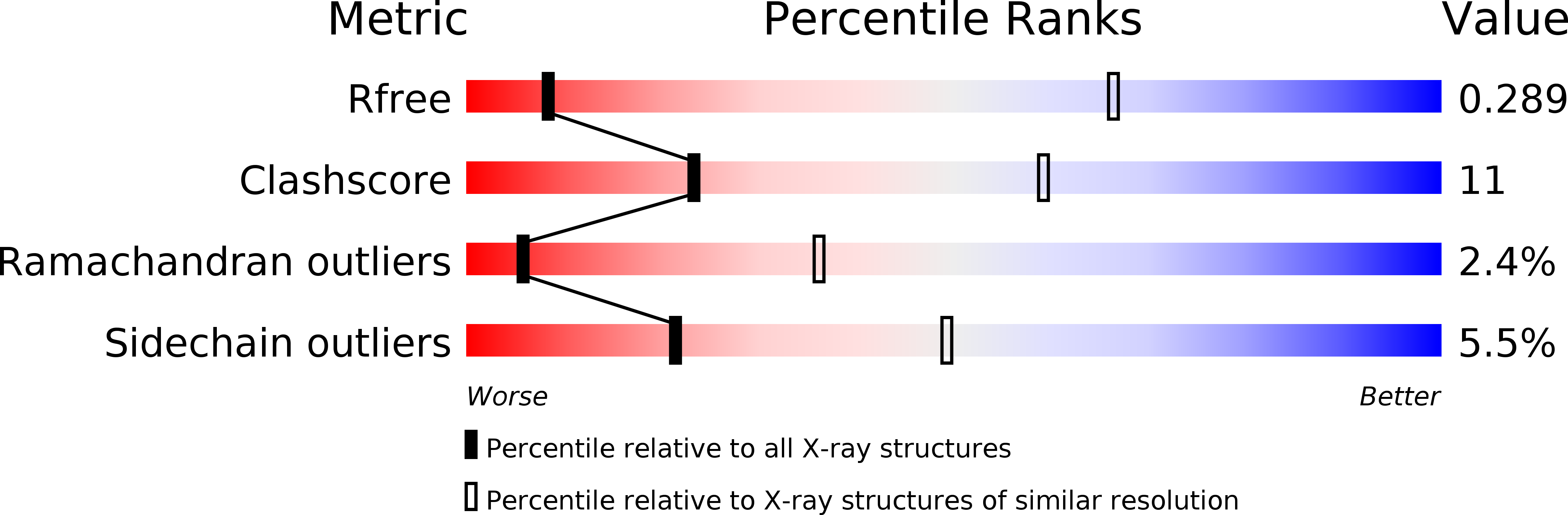
R-Value Free:
0.29
R-Value Work:
0.20
R-Value Observed:
0.21
Space Group:
P 21 21 21