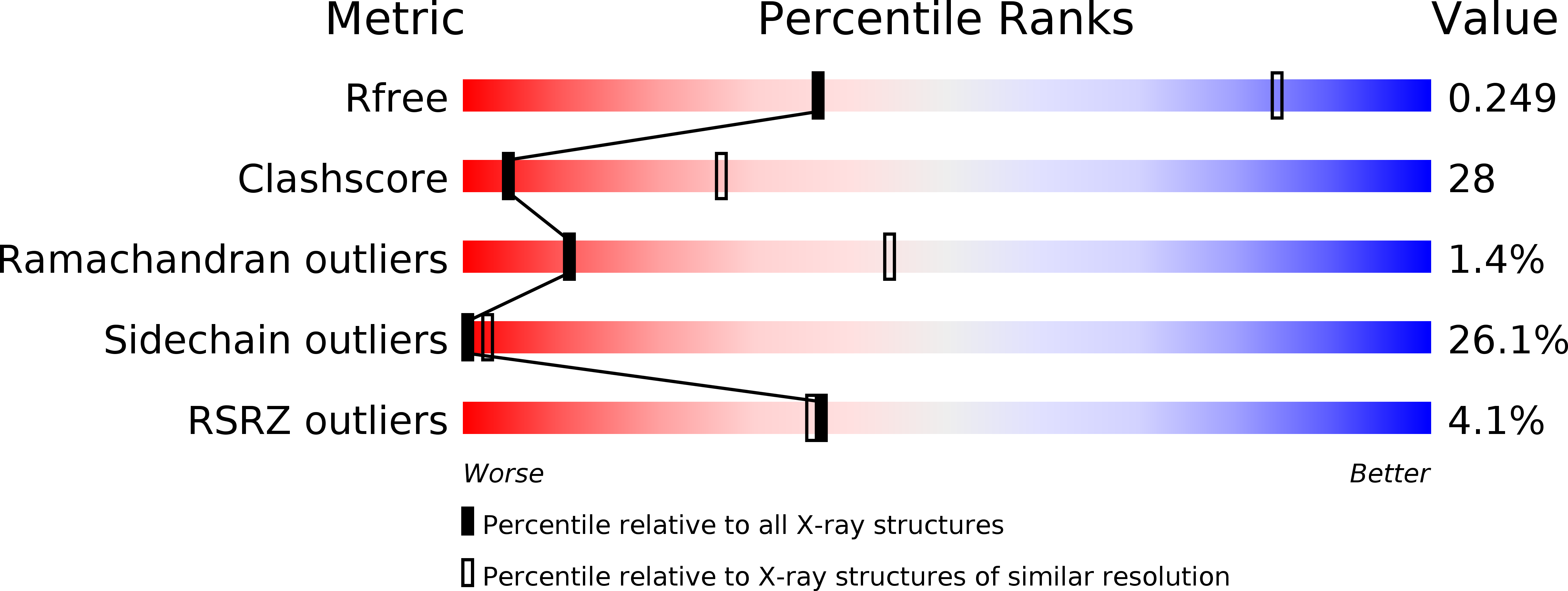
4QQV image
Deposition Date 2014-06-30
Release Date 2014-09-03
Last Version Date 2024-11-20
Entry Detail
PDB ID:
4QQV
Title:
Extracellular domains of mouse IL-3 beta receptor
Biological Source:
Source Organism:
Mus musculus (Taxon ID: 10090)
Host Organism:
Method Details:
Experimental Method:
Resolution:
3.45 Å
R-Value Free:
0.24
R-Value Work:
0.20
R-Value Observed:
0.20
Space Group:
C 1 2 1
Macromolecular Entities
Polymer Type:polypeptide(L)
Molecule:Interleukin-3 receptor class 2 subunit beta
Gene (Uniprot):Csf2rb2
Mutations:N328Q,K331A,R333A,D334A
Chain IDs:A, B, C, D
Chain Length:416
Number of Molecules:4
Biological Source:Mus musculus
Modified Residue
Compound ID Chain ID Parent Comp ID Details 2D Image
ASN C ASN GLYCOSYLATION SITE
Ligand Molecules
Primary Citation
Crystal structure of the mouse interleukin-3 beta-receptor: insights into interleukin-3 binding and receptor activation.
Biochem.J. 463 393 403 (2014)
PMID: 25137390 DOI: 10.1042/BJ20140863

Abstact

Interleukin-3 (IL-3) is a cytokine secreted by mast cells and activated T-cells known to be an important regulator of differentiation, survival, proliferation and activation of a range of haemopoietic lineages. The effects of IL-3 on target cells are mediated by a transmembrane receptor system composed of a cytokine-specific α-subunit and a β-subunit, the principal signalling entity. In the mouse, two β-subunits have co-evolved: a common β-subunit (βc) shared between IL-3 and the related cytokines IL-5 and granulocyte/macrophage colony-stimulating factor (GM-CSF); and an IL-3-specific β-subunit (βIL-3). βIL-3 differs from βc in its specificity for IL-3 and its capacity to bind IL-3 directly in the absence of an α-subunit, and, in the absence of structural information, the basis for these properties has remained enigmatic. In the present study, we have solved the crystal structure of the βIL-3 ectodomain at 3.45 Å (1 Å=0.1 nm) resolution. This structure provides the first evidence that βIL-3 adopts an arch-shaped intertwined homodimer with similar topology to the paralogous βc structure. In contrast with apo-βc, however, the ligand-binding interface of βIL-3 appears to pre-exist in a conformation receptive to IL-3 engagement. Molecular modelling of the IL-3-βIL-3 interface, in conjunction with previous mutational studies, suggests that divergent evolution of both βIL-3 and IL-3 underlies their unique capacity for direct interaction and specificity.

Legend

Protein

Chemical

Disease

Primary Citation of related structures