4QQ1 image
Deposition Date 2014-06-26
Release Date 2015-04-01
Last Version Date 2024-10-16
Entry Detail
PDB ID:
4QQ1
Keywords:
Title:
Crystal structure of the isotype 1 Transferrin binding protein B (TbpB) from serogroup B Neisseria meningitidis
Biological Source:
Source Organism(s):
Expression System(s):
Method Details:
Experimental Method:
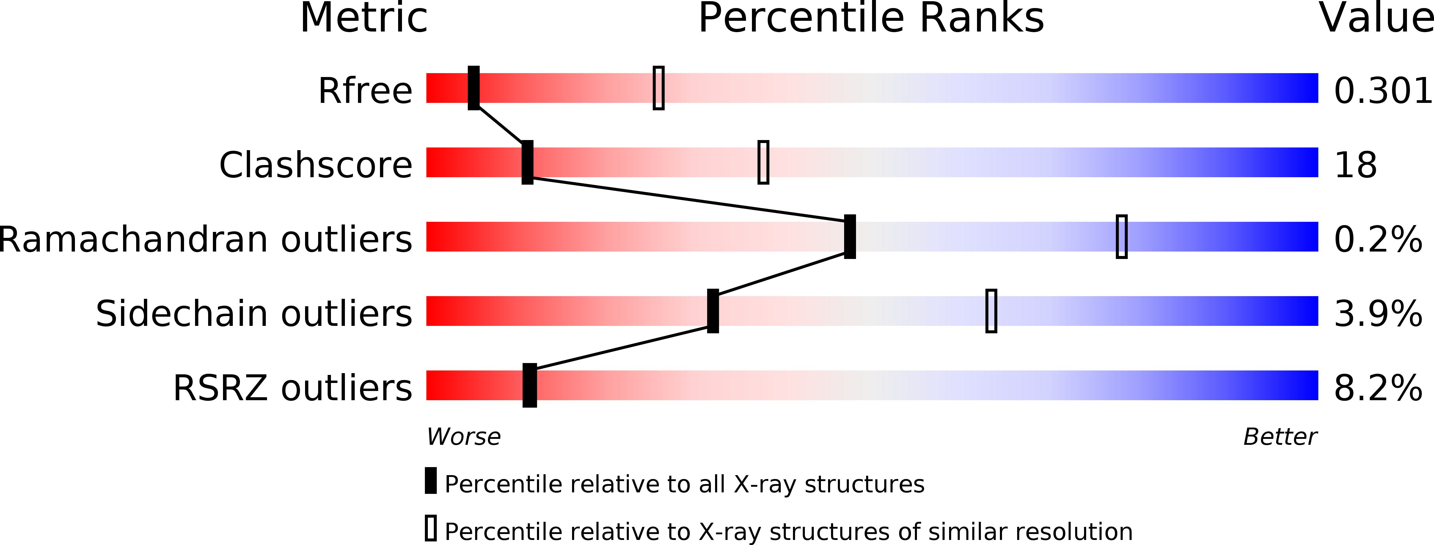
Resolution:
3.33 Å
R-Value Free:
0.30
R-Value Work:
0.24
R-Value Observed:
0.25
Space Group:
C 1 2 1
Macromolecular Entities
Structures with similar UniProt ID
Protein Blast
Polymer Type:polypeptide(L)
Molecule:Transferrin-binding protein 2
Gene (Uniprot):tbpB
Chain IDs:A, B, C
Chain Length:543
Number of Molecules:3
Biological Source:Neisseria meningitidis serogroup B
Primary Citation
Patterns of structural and sequence variation within isotype lineages of the Neisseria meningitidis transferrin receptor system.
Microbiologyopen 4 491 504 (2015)
PMID: 25800619 DOI: 10.1002/mbo3.254

Abstact

Neisseria meningitidis inhabits the human upper respiratory tract and is an important cause of sepsis and meningitis. A surface receptor comprised of transferrin-binding proteins A and B (TbpA and TbpB), is responsible for acquiring iron from host transferrin. Sequence and immunological diversity divides TbpBs into two distinct lineages; isotype I and isotype II. Two representative isotype I and II strains, B16B6 and M982, differ in their dependence on TbpB for in vitro growth on exogenous transferrin. The crystal structure of TbpB and a structural model for TbpA from the representative isotype I N. meningitidis strain B16B6 were obtained. The structures were integrated with a comprehensive analysis of the sequence diversity of these proteins to probe for potential functional differences. A distinct isotype I TbpA was identified that co-varied with TbpB and lacked sequence in the region for the loop 3 α-helix that is proposed to be involved in iron removal from transferrin. The tightly associated isotype I TbpBs had a distinct anchor peptide region, a distinct, smaller linker region between the lobes and lacked the large loops in the isotype II C-lobe. Sequences of the intact TbpB, the TbpB N-lobe, the TbpB C-lobe, and TbpA were subjected to phylogenetic analyses. The phylogenetic clustering of TbpA and the TbpB C-lobe were similar with two main branches comprising the isotype 1 and isotype 2 TbpBs, possibly suggesting an association between TbpA and the TbpB C-lobe. The intact TbpB and TbpB N-lobe had 4 main branches, one consisting of the isotype 1 TbpBs. One isotype 2 TbpB cluster appeared to consist of isotype 1 N-lobe sequences and isotype 2 C-lobe sequences, indicating the swapping of N-lobes and C-lobes. Our findings should inform future studies on the interaction between TbpB and TbpA and the process of iron acquisition.

Legend

Protein

Chemical

Disease

Primary Citation of related structures
Feedback Form
Name
Email
Institute
Feedback