4QPQ image
Entry Detail
PDB ID:
4QPQ
Title:
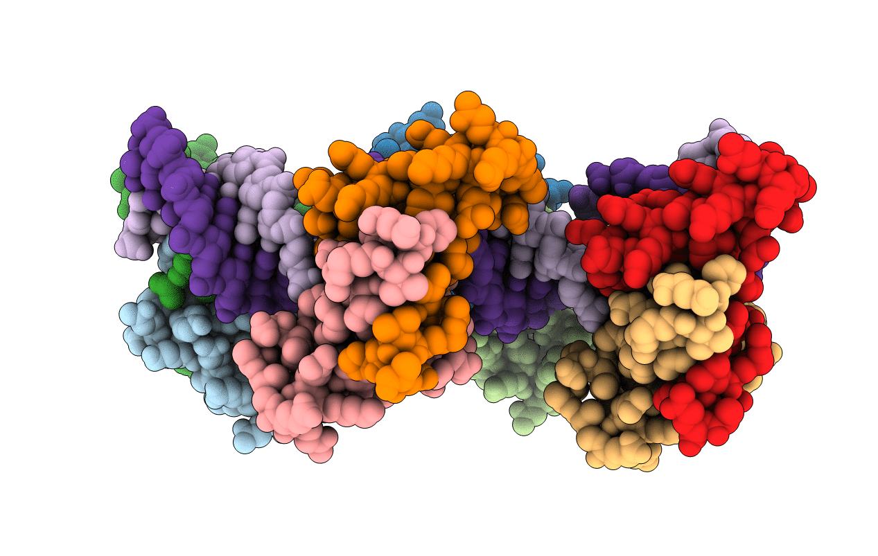
Mechanistic basis of plasmid-specific DNA binding of the F plasmid regulatory protein, TraM
Biological Source:
Source Organism:
Host Organism:
PDB Version:
Deposition Date:
2014-06-24
Release Date:
2014-09-24
Method Details:
Experimental Method:
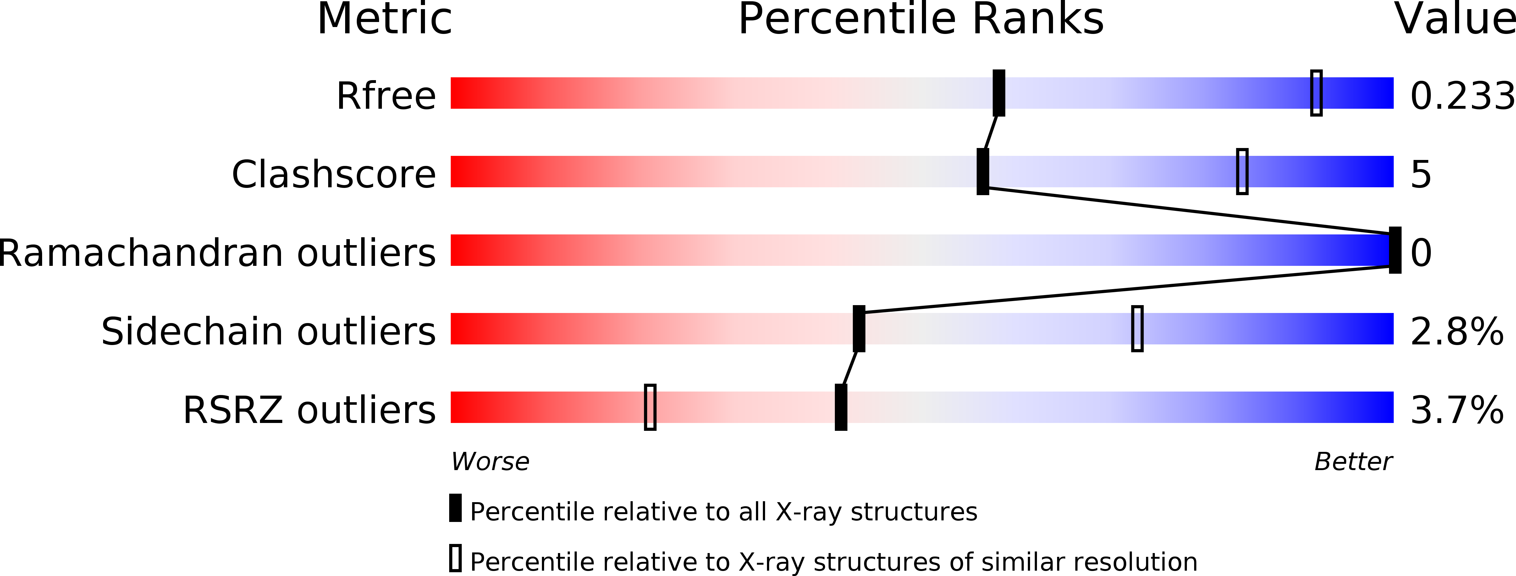
Resolution:
3.11 Å
R-Value Free:
0.23
R-Value Work:
0.22
R-Value Observed:
0.22
Space Group:
P 43 21 2
Macromolecular Entities
Polymer Type:polypeptide(L)
Description:Relaxosome protein TraM
Chain IDs:A, B, C, D, E, F, G, H
Chain Length:53
Number of Molecules:8
Biological Source:Escherichia coli
Polymer Type:polydeoxyribonucleotide
Description:sbmA DNA1
Chain IDs:I (auth: P)
Chain Length:28
Number of Molecules:1
Biological Source:
Polymer Type:polydeoxyribonucleotide
Description:sbmA DNA2
Chain IDs:J (auth: Q)
Chain Length:28
Number of Molecules:1
Biological Source:
Ligand Molecules
Primary Citation
Mechanistic Basis of Plasmid-Specific DNA Binding of the F Plasmid Regulatory Protein, TraM.
J.Mol.Biol. 426 3783 3795 (2014)
PMID: 25284757 DOI: 10.1016/j.jmb.2014.09.018

Abstact

The conjugative transfer of bacterial F plasmids relies on TraM, a plasmid-encoded protein that recognizes multiple DNA sites to recruit the plasmid to the conjugative pore. In spite of the high degree of amino acid sequence conservation between TraM proteins, many of these proteins have markedly different DNA binding specificities that ensure the selective recruitment of a plasmid to its cognate pore. Here we present the structure of F TraM RHH (ribbon-helix-helix) domain bound to its sbmA site. The structure indicates that a pair of TraM tetramers cooperatively binds an underwound sbmA site containing 12 base pairs per turn. The sbmA is composed of 4 copies of a 5-base-pair motif, each of which is recognized by an RHH domain. The structure reveals that a single conservative amino acid difference in the RHH β-ribbon between F and pED208 TraM changes its specificity for its cognate 5-base-pair sequence motif. Specificity is also dictated by the positioning of 2-base-pair spacer elements within sbmA; in F sbmA, the spacers are positioned between motifs 1 and 2 and between motifs 3 and 4, whereas in pED208 sbmA, there is a single spacer between motifs 2 and 3. We also demonstrate that a pair of F TraM tetramers can cooperatively bind its sbmC site with an affinity similar to that of sbmA in spite of a lack of sequence similarity between these DNA elements. These results provide a basis for the prediction of the DNA binding properties of the family of TraM proteins.

Legend

Protein

Chemical

Disease

Primary Citation of related structures