4QPB image
Deposition Date 2014-06-22
Release Date 2014-07-09
Last Version Date 2023-09-20
Entry Detail
PDB ID:
4QPB
Keywords:
Title:
Catalytic domain of the antimicrobial peptidase lysostaphin from Staphylococcus simulans crystallized in the absence of phosphate
Biological Source:
Source Organism:
Host Organism:
Method Details:
Experimental Method:
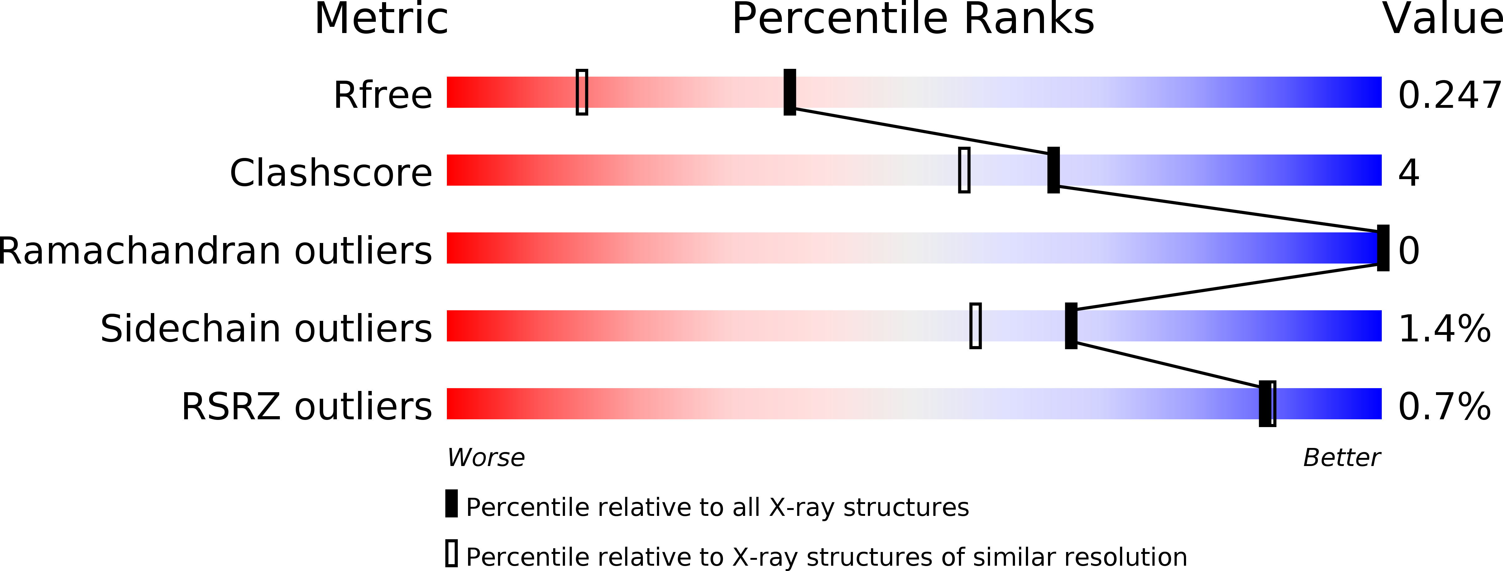
Resolution:
1.78 Å
R-Value Free:
0.24
R-Value Work:
0.21
R-Value Observed:
0.21
Space Group:
P 1 21 1
Macromolecular Entities
Polymer Type:polypeptide(L)
Molecule:Lysostaphin
Gene (Uniprot):lss
Chain IDs:A, B
Chain Length:140
Number of Molecules:2
Biological Source:Staphylococcus simulans
Primary Citation
Crystal structure of the antimicrobial peptidase lysostaphin from Staphylococcus simulans.
Febs J. 281 4112 4122 (2014)
PMID: 25039253 DOI: 10.1111/febs.12929

Abstact

UNLABELLED Staphylococcus simulans biovar staphylolyticus lysostaphin efficiently cleaves Staphylococcus aureus cell walls. The protein is in late clinical trials as a topical anti-staphylococcal agent, and can be used to prevent staphylococcal growth on artificial surfaces. Moreover, the gene has been both stably engineered into and virally delivered to mice or livestock to obtain resistance against staphylococci. Here, we report the first crystal structure of mature lysostaphin and two structures of its isolated catalytic domain at 3.5, 1.78 and 1.26 Å resolution, respectively. The structure of the mature active enzyme confirms its expected organization into catalytic and cell-wall-targeting domains. It also indicates that the domains are mobile with respect to each other because of the presence of a highly flexible peptide linker. The high-resolution structures of the catalytic domain provide details of Zn(2+) coordination and may serve as a starting point for the engineering of lysostaphin variants with improved biotechnological characteristics. STRUCTURED DIGITAL ABSTRACT lysostaphin by x-ray crystallography (1, 2).

Legend

Protein

Chemical

Disease

Primary Citation of related structures