4QMK image
Deposition Date 2014-06-16
Release Date 2014-12-17
Last Version Date 2023-09-20
Entry Detail
PDB ID:
4QMK
Keywords:
Title:
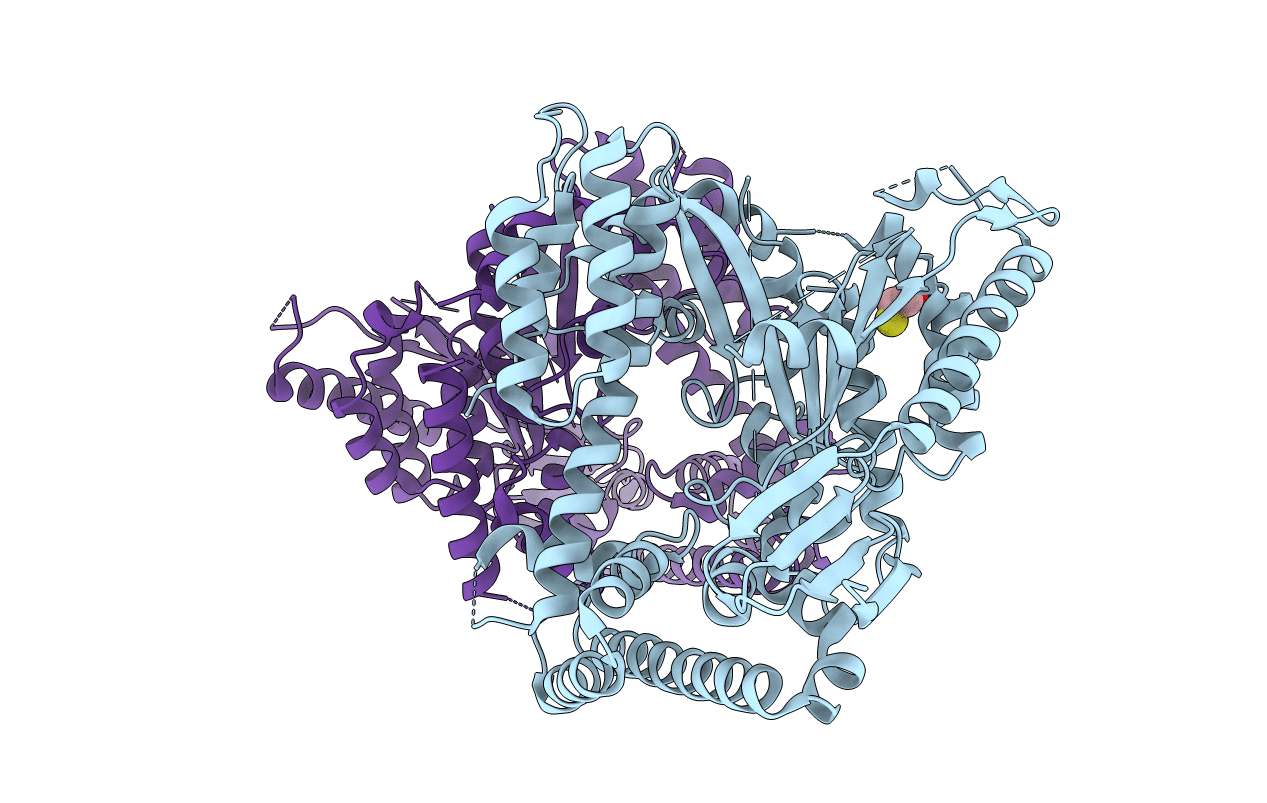
Crystal structure of type III effector protein ExoU (exoU)
Biological Source:
Source Organism:
Host Organism:
Method Details:
Experimental Method:
Resolution:
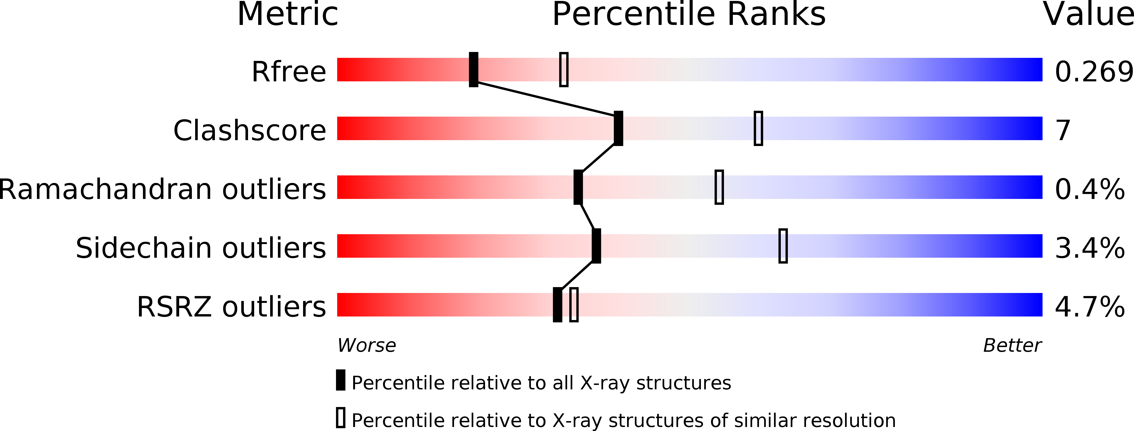
2.50 Å
R-Value Free:
0.26
R-Value Work:
0.20
R-Value Observed:
0.20
Space Group:
P 1 21 1
Macromolecular Entities
Polymer Type:polypeptide(L)
Molecule:Type III secretion system effector protein ExoU
Chain IDs:A, B
Chain Length:677
Number of Molecules:2
Biological Source:Pseudomonas fluorescens
Ligand Molecules
Primary Citation
A Novel Phosphatidylinositol 4,5-Bisphosphate Binding Domain Mediates Plasma Membrane Localization of ExoU and Other Patatin-like Phospholipases.
J.Biol.Chem. 290 2919 2937 (2015)
PMID: 25505182 DOI: 10.1074/jbc.M114.611251

Abstact

Bacterial toxins require localization to specific intracellular compartments following injection into host cells. In this study, we examined the membrane targeting of a broad family of bacterial proteins, the patatin-like phospholipases. The best characterized member of this family is ExoU, an effector of the Pseudomonas aeruginosa type III secretion system. Upon injection into host cells, ExoU localizes to the plasma membrane, where it uses its phospholipase A2 activity to lyse infected cells. The targeting mechanism of ExoU is poorly characterized, but it was recently found to bind to the phospholipid phosphatidylinositol 4,5-bisphosphate (PI(4,5)P2), a marker for the plasma membrane of eukaryotic cells. We confirmed that the membrane localization domain (MLD) of ExoU had a direct affinity for PI(4,5)P2, and we determined that this binding was required for ExoU localization. Previously uncharacterized ExoU homologs from Pseudomonas fluorescens and Photorhabdus asymbiotica also localized to the plasma membrane and required PI(4,5)P2 for this localization. A conserved arginine within the MLD was critical for interaction of each protein with PI(4,5)P2 and for localization. Furthermore, we determined the crystal structure of the full-length P. fluorescens ExoU and found that it was similar to that of P. aeruginosa ExoU. Each MLD contains a four-helical bundle, with the conserved arginine exposed at its cap to allow for interaction with the negatively charged PI(4,5)P2. Overall, these findings provide a structural explanation for the targeting of patatin-like phospholipases to the plasma membrane and define the MLD of ExoU as a member of a new class of PI(4,5)P2 binding domains.

Legend

Protein

Chemical

Disease

Primary Citation of related structures