4QKW image
Deposition Date 2014-06-10
Release Date 2015-03-18
Last Version Date 2024-03-20
Entry Detail
PDB ID:
4QKW
Title:
Crystal structure of the zebrafish cavin4a HR1 domain
Biological Source:
Source Organism:
Danio rerio (Taxon ID: 7955)
Host Organism:
Method Details:
Experimental Method:
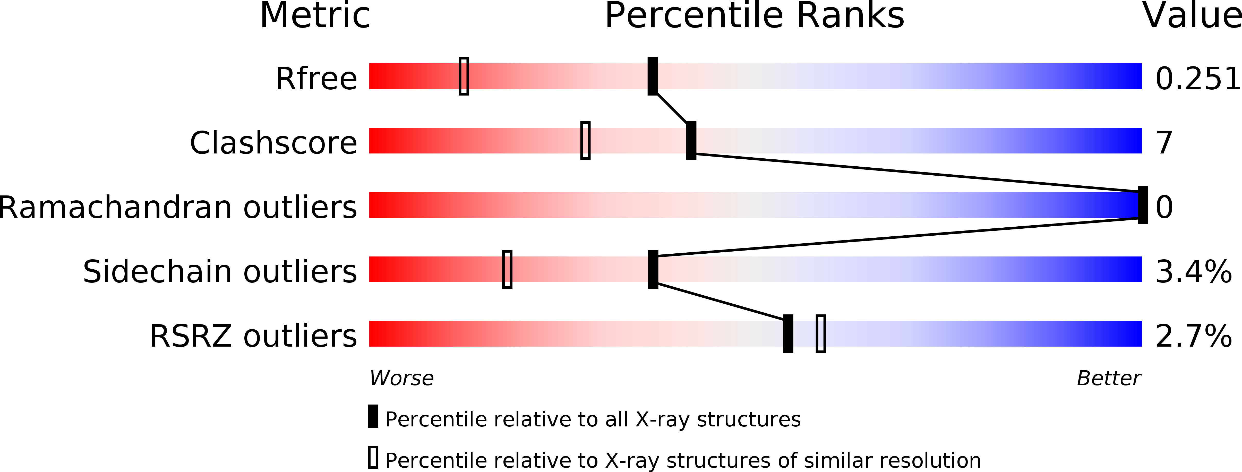
Resolution:
1.70 Å
R-Value Free:
0.24
R-Value Work:
0.21
R-Value Observed:
0.21
Space Group:
C 1 2 1
Macromolecular Entities
Polymer Type:polypeptide(L)
Molecule:Muscle-related coiled-coil protein
Gene (Uniprot):cavin4a
Chain IDs:A, B, C
Chain Length:108
Number of Molecules:3
Biological Source:Danio rerio
Ligand Molecules
Primary Citation
Structural insights into the organization of the cavin membrane coat complex
Dev.Cell 31 405 419 (2014)
PMID: 25453557 DOI: 10.1016/j.devcel.2014.10.002

Abstact

Caveolae are cell-surface membrane invaginations that play critical roles in cellular processes including signaling and membrane homeostasis. The cavin proteins, in cooperation with caveolins, are essential for caveola formation. Here we show that a minimal N-terminal domain of the cavins, termed HR1, is required and sufficient for their homo- and hetero-oligomerization. Crystal structures of the mouse cavin1 and zebrafish cavin4a HR1 domains reveal highly conserved trimeric coiled-coil architectures, with intersubunit interactions that determine the specificity of cavin-cavin interactions. The HR1 domain contains a basic surface patch that interacts with polyphosphoinositides and coordinates with additional membrane-binding sites within the cavin C terminus to facilitate membrane association and remodeling. Electron microscopy of purified cavins reveals the existence of large assemblies, composed of a repeating rod-like structural element, and we propose that these structures polymerize through membrane-coupled interactions to form the unique striations observed on the surface of caveolae in vivo.

Legend

Protein

Chemical

Disease

Primary Citation of related structures