4QI2 image
Entry Detail
PDB ID:
4QI2
Title:
X-ray structure of the ROQ domain from murine Roquin-1 in complex with a 23-mer Tnf-CDE RNA
Biological Source:
Source Organism:
Host Organism:
PDB Version:
Deposition Date:
2014-05-30
Release Date:
2014-07-16
Method Details:
Experimental Method:
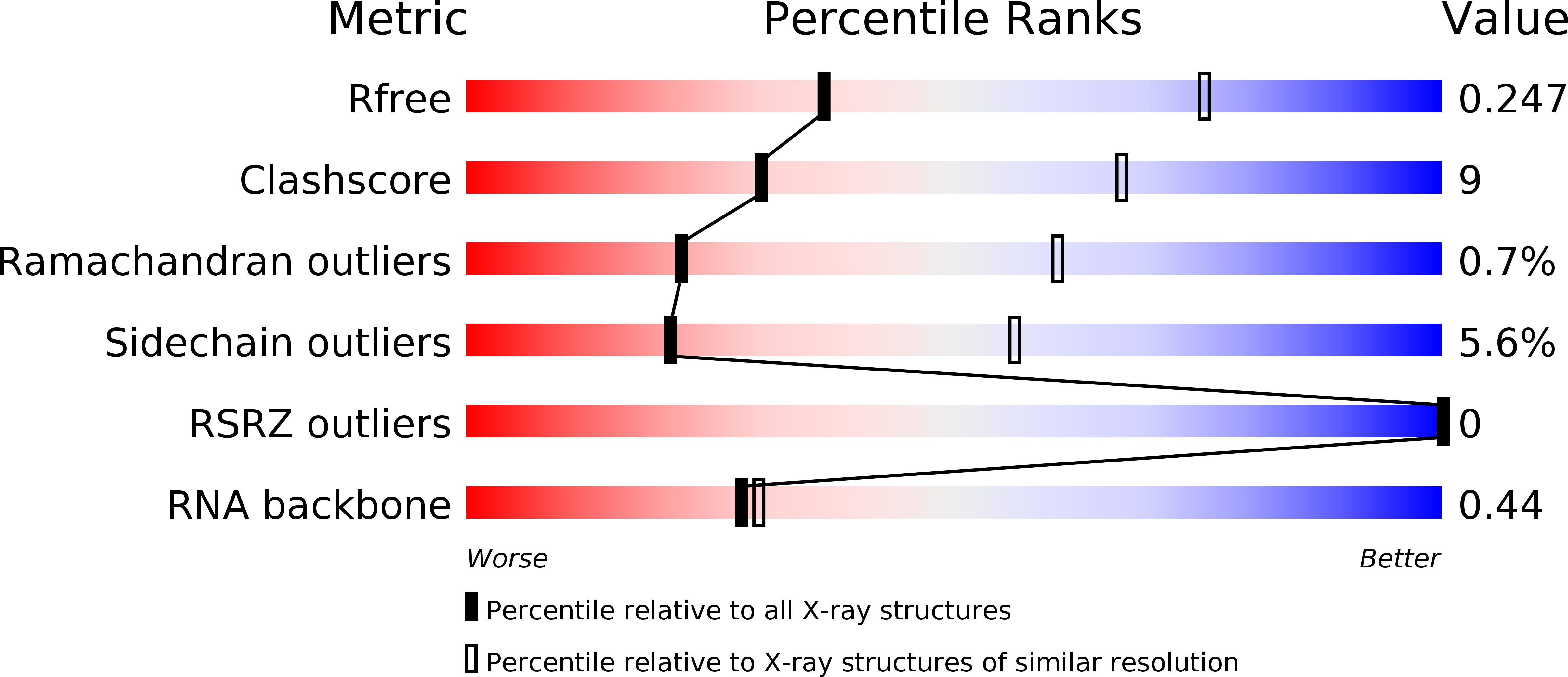
Resolution:
3.00 Å
R-Value Free:
0.24
R-Value Work:
0.20
R-Value Observed:
0.20
Space Group:
P 1
Macromolecular Entities
Polymer Type:polypeptide(L)
Description:Roquin-1
Chain IDs:A, B, C, D
Chain Length:180
Number of Molecules:4
Biological Source:Mus musculus
Polymer Type:polyribonucleotide
Description:RNA (5'-R(*AP*CP*AP*UP*GP*UP*UP*UP*UP*CP*UP*GP*UP*GP*AP*AP*AP*AP*CP*GP*GP*AP*G)-3')
Chain IDs:E, F, G, H
Chain Length:23
Number of Molecules:4
Biological Source:
Primary Citation
Structural basis for RNA recognition in roquin-mediated post-transcriptional gene regulation.
Nat.Struct.Mol.Biol. 21 671 678 (2014)
PMID: 25026077 DOI: 10.1038/nsmb.2855

Abstact

Roquin function in T cells is essential for the prevention of autoimmune disease. Roquin interacts with the 3' untranslated regions (UTRs) of co-stimulatory receptors and controls T-cell activation and differentiation. Here we show that the N-terminal ROQ domain from mouse roquin adopts an extended winged-helix (WH) fold, which is sufficient for binding to the constitutive decay element (CDE) in the Tnf 3' UTR. The crystal structure of the ROQ domain in complex with a prototypical CDE RNA stem-loop reveals tight recognition of the RNA stem and its triloop. Surprisingly, roquin uses mainly non-sequence-specific contacts to the RNA, thus suggesting a relaxed CDE consensus and implicating a broader spectrum of target mRNAs than previously anticipated. Consistently with this, NMR and binding experiments with CDE-like stem-loops together with cell-based assays confirm roquin-dependent regulation of relaxed CDE consensus motifs in natural 3' UTRs.

Legend

Protein

Chemical

Disease

Primary Citation of related structures