4QGX image
Deposition Date 2014-05-26
Release Date 2015-01-28
Last Version Date 2024-10-09
Entry Detail
PDB ID:
4QGX
Keywords:
Title:
Crystal structure of the R132K:R111L:L121E mutant of Cellular Retinoic Acid Binding ProteinII complexed with a synthetic ligand (Merocyanine) at 1.47 angstrom resolution
Biological Source:
Source Organism:
Homo sapiens (Taxon ID: 9606)
Host Organism:
Method Details:
Experimental Method:
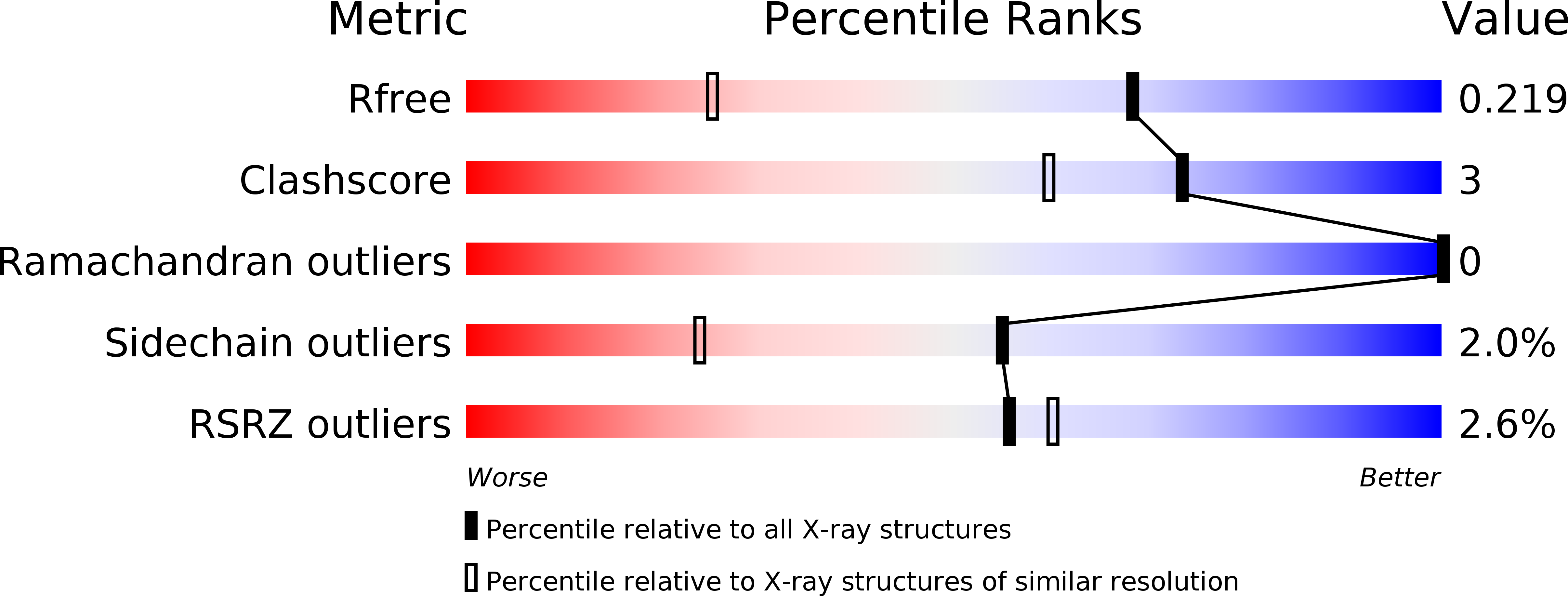
Resolution:
1.47 Å
R-Value Free:
0.22
R-Value Work:
0.18
R-Value Observed:
0.19
Space Group:
P 1
Macromolecular Entities
Polymer Type:polypeptide(L)
Molecule:Cellular retinoic acid-binding protein 2
Gene (Uniprot):CRABP2
Mutations:R132K, R111L, L121E
Chain IDs:A, B
Chain Length:137
Number of Molecules:2
Biological Source:Homo sapiens
Ligand Molecules
Primary Citation
"Turn-on" protein fluorescence: in situ formation of cyanine dyes.
J.Am.Chem.Soc. 137 1073 1080 (2015)
PMID: 25534273 DOI: 10.1021/ja506376j

Abstact

Protein reengineering of cellular retinoic acid binding protein II (CRABPII) has yielded a genetically addressable system, capable of binding a profluorophoric chromophore that results in fluorescent protein/chromophore complexes. These complexes exhibit far-red emission, with high quantum efficiencies and brightness and also exhibit excellent pH stability spanning the range of 2-11. In the course of this study, it became evident that single mutations of L121E and R59W were most effective in improving the fluorescent characteristics of CRABPII mutants as well as the kinetics of complex formation. The readily crystallizable nature of these proteins was invaluable to provide clues for the observed spectroscopic behavior that results from single mutation of key residues.

Legend

Protein

Chemical

Disease

Primary Citation of related structures