4QF9 image
Deposition Date 2014-05-20
Release Date 2015-04-22
Last Version Date 2023-09-20
Entry Detail
PDB ID:
4QF9
Title:
Structure of GluK1 ligand-binding domain (S1S2) in complex with (S)-2-amino-4-(2,3-dioxo-1,2,3,4-tetrahydroquinoxalin-6-yl)butanoic acid at 2.28 A resolution
Biological Source:
Source Organism:
Host Organism:
Method Details:
Experimental Method:
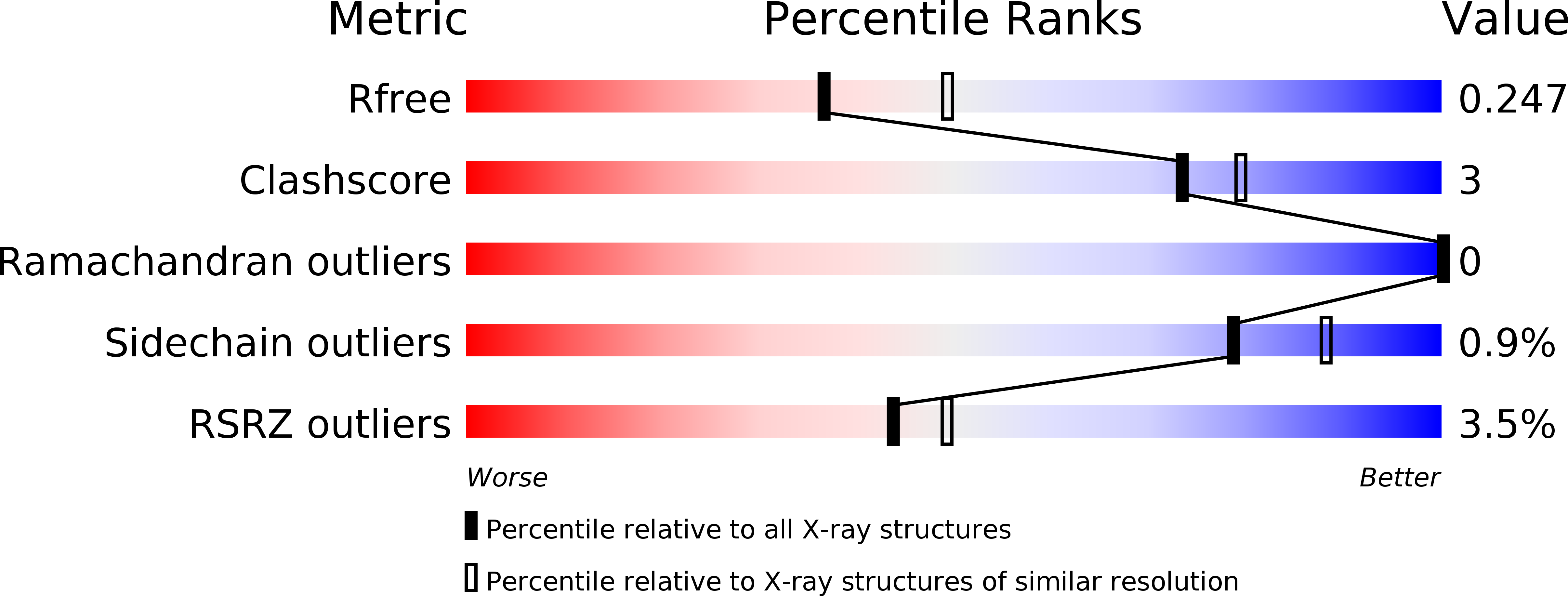
Resolution:
2.28 Å
R-Value Free:
0.24
R-Value Work:
0.19
R-Value Observed:
0.20
Space Group:
C 1 2 1
Macromolecular Entities
Polymer Type:polypeptide(L)
Molecule:Glutamate receptor ionotropic, kainate 1
Gene (Uniprot):Grik1
Chain IDs:A, B, C
Chain Length:257
Number of Molecules:3
Biological Source:Rattus norvegicus
Primary Citation
Binding Mode of an alpha-Amino Acid-Linked Quinoxaline-2,3-dione Analogue at Glutamate Receptor Subtype GluK1.
ACS Chem Neurosci 6 845 854 (2015)
PMID: 25856736 DOI: 10.1021/acschemneuro.5b00038

Abstact

Two α-amino acid-functionalized quinoxalines, 1a (CNG-10301) and 1b (CNG-10300), of a quinoxaline moiety coupled to an amino acid moiety were designed, synthesized, and characterized pharmacologically. While 1a displayed low affinity at native AMPA, KA, and NMDA receptors, and at homomeric GluK1,3 receptors, the affinity for GluK2 was in the midmicromolar range (Ki = 136 μM), 1b displayed low to midmicromolar range binding affinity at all the iGluRs (Ki = 9-126 μM). In functional experiments (outside-out patches excised from transfected HEK293T cells), 100 μM 1a partially blocked GluK1 (33% peak response), while GluK2 was unaffected (96% peak response). Furthermore, 1a was shown not to be an agonist at GluK1 and GluK2 at 100 μM. On the other hand, 100 μM 1b fully antagonized GluK1 (8% peak response) but only partially blocked GluK2 (33% peak response). An X-ray structure at 2.3 Å resolution of 1b in the GluK1-LBD (ligand-binding domain) disclosed an unexpected binding mode compared to the predictions made during the design phase; the quinoxaline moiety remains to act as an amino acid bioisostere, but the amino acid moiety is oriented into a new area within the GluK1 receptor. The structure of the GluK1-LBD with 1b showed a large variation in domain openings of the three molecules from 25° to 49°, demonstrating that the GluK1-LBD is capable of undergoing major domain movements.

Legend

Protein

Chemical

Disease

Primary Citation of related structures