4QDD image
Entry Detail
PDB ID:
4QDD
Title:
Crystal structure of 3-ketosteroid-9-alpha-hydroxylase 5 (KshA5) from R. rhodochrous in complex with 1,4-30Q-CoA
Biological Source:
Source Organism:
Host Organism:
PDB Version:
Deposition Date:
2014-05-13
Release Date:
2014-07-30
Method Details:
Experimental Method:
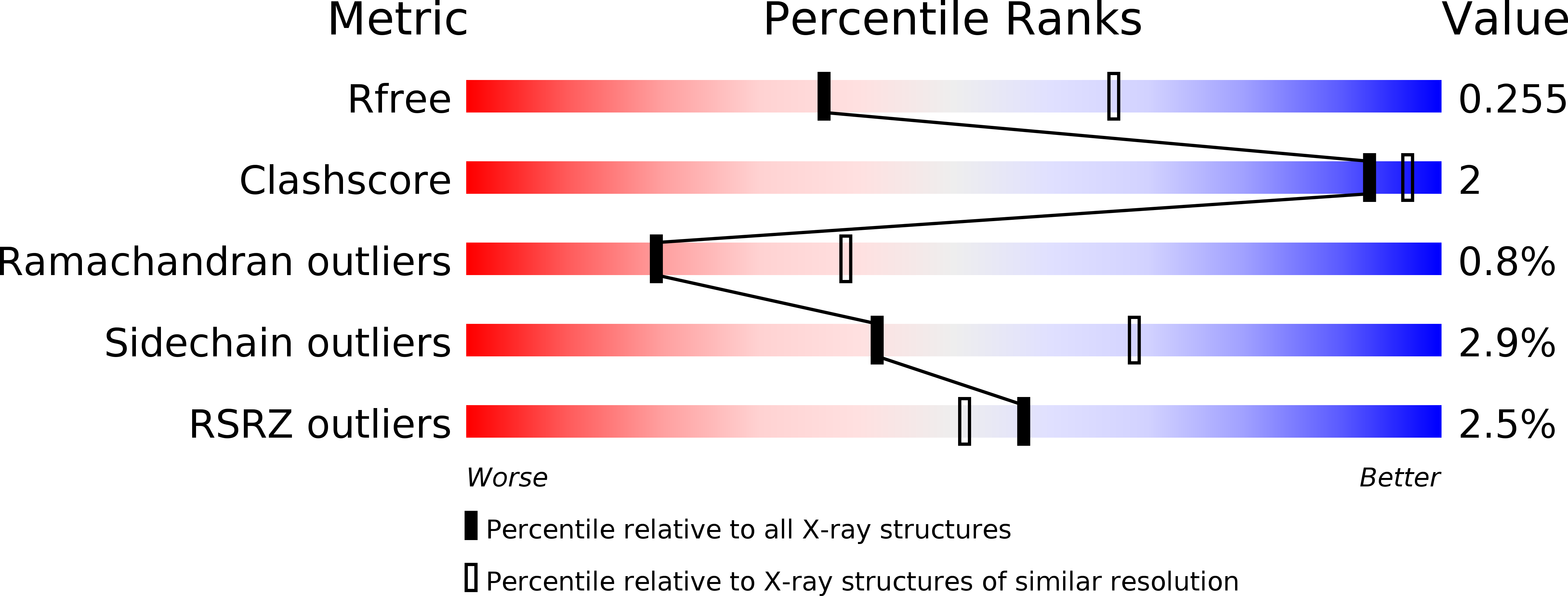
Resolution:
2.60 Å
R-Value Free:
0.25
R-Value Work:
0.22
R-Value Observed:
0.22
Space Group:
P 63
Macromolecular Entities
Polymer Type:polypeptide(L)
Description:3-ketosteroid 9alpha-hydroxylase oxygenase
Chain IDs:A
Chain Length:390
Number of Molecules:1
Biological Source:Rhodococcus rhodochrous
Primary Citation
Substrate specificities and conformational flexibility of 3-ketosteroid 9 alpha-hydroxylases.
J.Biol.Chem. 289 25523 25536 (2014)
PMID: 25049233 DOI: 10.1074/jbc.M114.575886

Abstact

KshA is the oxygenase component of 3-ketosteroid 9α-hydroxylase, a Rieske oxygenase involved in the bacterial degradation of steroids. Consistent with its role in bile acid catabolism, KshA1 from Rhodococcus rhodochrous DSM43269 had the highest apparent specificity (kcat/Km) for steroids with an isopropyl side chain at C17, such as 3-oxo-23,24-bisnorcholesta-1,4-diene-22-oate (1,4-BNC). By contrast, the KshA5 homolog had the highest apparent specificity for substrates with no C17 side chain (kcat/Km >10(5) s(-1) M(-1) for 4-estrendione, 5α-androstandione, and testosterone). Unexpectedly, substrates such as 4-androstene-3,17-dione (ADD) and 4-BNC displayed strong substrate inhibition (Ki S ∼100 μM). By comparison, the cholesterol-degrading KshAMtb from Mycobacterium tuberculosis had the highest specificity for CoA-thioesterified substrates. These specificities are consistent with differences in the catabolism of cholesterol and bile acids, respectively, in actinobacteria. X-ray crystallographic structures of the KshAMtb·ADD, KshA1·1,4-BNC-CoA, KshA5·ADD, and KshA5·1,4-BNC-CoA complexes revealed that the enzymes have very similar steroid-binding pockets with the substrate's C17 oriented toward the active site opening. Comparisons suggest Tyr-245 and Phe-297 are determinants of KshA1 specificity. All enzymes have a flexible 16-residue "mouth loop," which in some structures completely occluded the substrate-binding pocket from the bulk solvent. Remarkably, the catalytic iron and α-helices harboring its ligands were displaced up to 4.4 Å in the KshA5·substrate complexes as compared with substrate-free KshA, suggesting that Rieske oxygenases may have a dynamic nature similar to cytochrome P450.

Legend

Protein

Chemical

Disease

Primary Citation of related structures