4QCA image
Deposition Date 2014-05-09
Release Date 2015-05-13
Last Version Date 2023-09-20
Entry Detail
PDB ID:
4QCA
Keywords:
Title:
Crystal structure of Vaccinia virus uracil-DNA glycosylase mutant R167AD4
Biological Source:
Source Organism:
Host Organism:
Method Details:
Experimental Method:
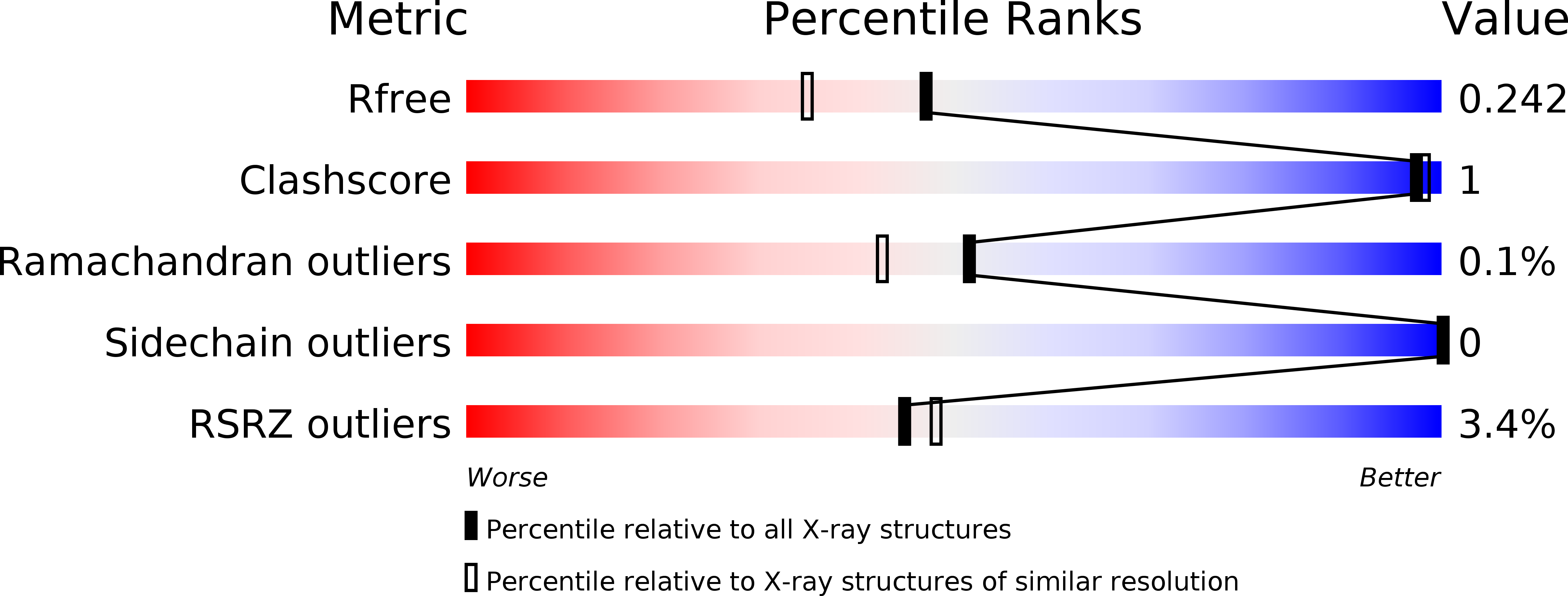
Resolution:
1.90 Å
R-Value Free:
0.23
R-Value Work:
0.20
R-Value Observed:
0.20
Space Group:
P 21 21 21
Macromolecular Entities
Polymer Type:polypeptide(L)
Molecule:Uracil-DNA glycosylase
Mutations:D17N, R167A
Chain IDs:A, B, C, D
Chain Length:221
Number of Molecules:4
Biological Source:Vaccinia virus
Primary Citation
Crystallization and preliminary X-ray diffraction analysis of three recombinant mutants of Vaccinia virus uracil DNA glycosylase.
Acta Crystallogr.,Sect.F 69 295 301 (2013)
PMID: 23519808 DOI: 10.1107/S1744309113002716

Abstact

Amino-acid residues located at a highly flexible area in the uracil DNA glycosylase of Vaccinia virus were mutated. In the crystal structure of wild-type D4 these residues lie at the dimer interface. Specifically, three mutants were generated: (i) residue Arg167 was replaced with an alanine (R167AD4), (ii) residues Glu171, Ser172 and Pro173 were substituted with three glycine residues (3GD4) and (iii) residues Glu171 and Ser172 were deleted (Δ171-172D4). Mutant proteins were expressed, purified and crystallized in order to investigate the effects of these mutations on the structure of the protein.

Legend

Protein

Chemical

Disease

Primary Citation of related structures