4QAZ image
Deposition Date 2014-05-06
Release Date 2014-09-10
Last Version Date 2024-02-28
Entry Detail
PDB ID:
4QAZ
Keywords:
Title:
The crystal structure of the C-terminal domain of Ebola (Zaire) nucleoprotein
Biological Source:
Source Organism:
Ebola virus (Taxon ID: 128952)
Host Organism:
Method Details:
Experimental Method:
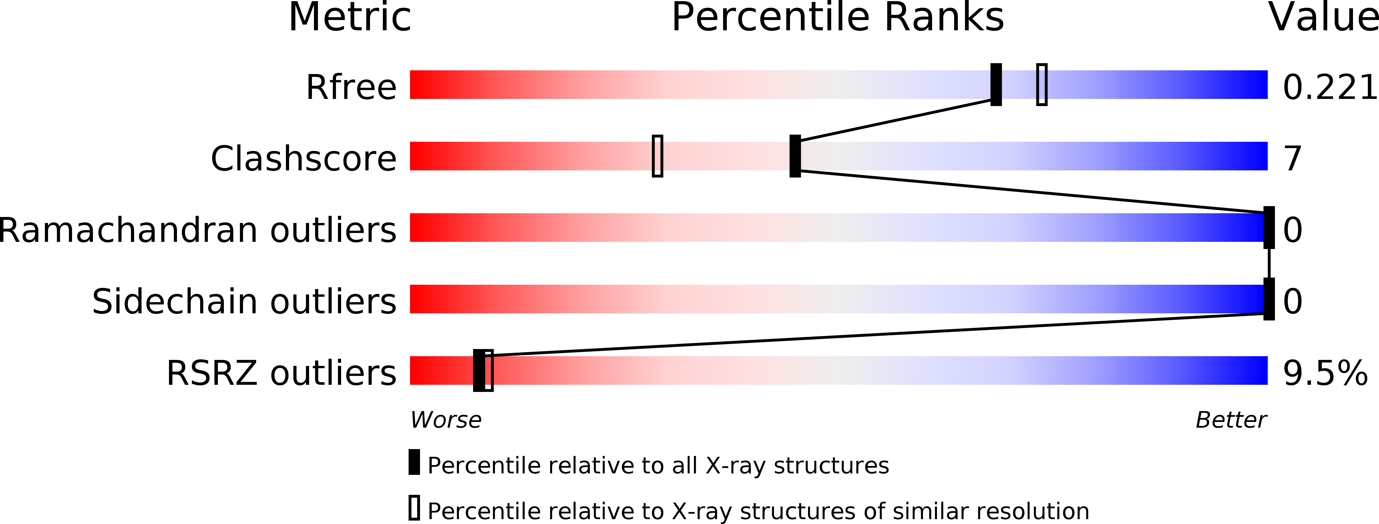
Resolution:
1.98 Å
R-Value Free:
0.22
R-Value Work:
0.18
R-Value Observed:
0.18
Space Group:
P 31 2 1
Macromolecular Entities
Polymer Type:polypeptide(L)
Molecule:Nucleoprotein
Gene (Uniprot):NP
Chain IDs:A
Chain Length:103
Number of Molecules:1
Biological Source:Ebola virus
Primary Citation
The structure of the C-terminal domain of the Zaire ebolavirus nucleoprotein.
Acta Crystallogr.,Sect.D 70 2420 2429 (2014)
PMID: 25195755 DOI: 10.1107/S1399004714014710

Abstact

Ebolavirus (EBOV) causes severe hemorrhagic fever with a mortality rate of up to 90%. EBOV is a member of the order Mononegavirales and, like other viruses in this taxonomic group, contains a negative-sense single-stranded (ss) RNA. The EBOV ssRNA encodes seven distinct proteins. One of them, the nucleoprotein (NP), is the most abundant viral protein in the infected cell and within the viral nucleocapsid. Like other EBOV proteins, NP is multifunctional. It is tightly associated with the viral genome and is essential for viral transcription, RNA replication, genome packaging and nucleocapsid assembly prior to membrane encapsulation. NP is unusual among the Mononegavirales in that it contains two distinct regions, or putative domains, the C-terminal of which shows no homology to any known proteins and is purported to be a hub for protein-protein interactions within the nucleocapsid. The atomic structure of NP remains unknown. Here, the boundaries of the N- and C-terminal domains of NP from Zaire EBOV are defined, it is shown that they can be expressed as highly stable recombinant proteins in Escherichia coli, and the atomic structure of the C-terminal domain (residues 641-739) derived from analysis of two distinct crystal forms at 1.98 and 1.75 Å resolution is described. The structure reveals a novel tertiary fold that is distantly reminiscent of the β-grasp architecture.

Legend

Protein

Chemical

Disease

Primary Citation of related structures