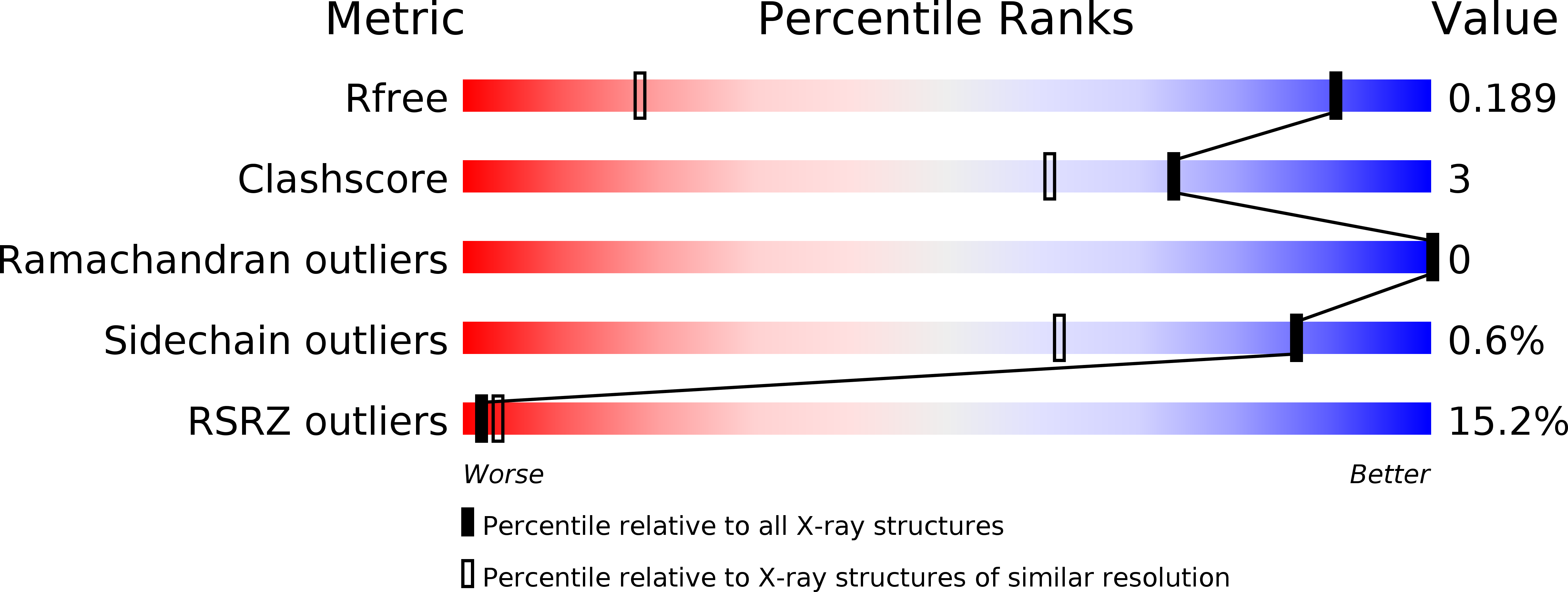
4QA8 image
Deposition Date 2014-05-02
Release Date 2014-10-22
Last Version Date 2024-03-20
Entry Detail
PDB ID:
4QA8
Keywords:
Title:
Crystal structure of LprF from Mycobacterium bovis
Biological Source:
Source Organism:
Host Organism:
Method Details:
Experimental Method:
Resolution:
1.10 Å
R-Value Free:
0.18
R-Value Work:
0.16
R-Value Observed:
0.16
Space Group:
P 1
Macromolecular Entities
Polymer Type:polypeptide(L)
Molecule:Putative lipoprotein LprF
Gene (Uniprot):lprF
Chain IDs:A
Chain Length:229
Number of Molecules:1
Biological Source:Mycobacterium bovis
Ligand Molecules
Primary Citation
Crystal structure and functional implications of LprF from Mycobacterium tuberculosis and M. bovis
Acta Crystallogr.,Sect.D 70 2619 2630 (2014)
PMID: 25286846 DOI: 10.1107/S1399004714016599

Abstact

The Gram-positive bacteria Mycobacterium tuberculosis and M. bovis are causative agents of tuberculosis in humans and cattle. The lipoprotein LprF is found in M. tuberculosis and M. bovis but not in the nonpathogenic M. smegmatis. To date, the role of LprF remains to be elucidated. In this study, the crystal structure of LprF has been determined at 1.1 Å resolution. The overall structure is similar to that of a homologue, LprG, with a central hydrophobic cavity that binds a triacylated glycolipid. LprF exhibited a central cavity structure similar to that of LprG, but with a smaller cavity that binds two alkyl chains. Consistently, subsequent mass-spectrometric analysis revealed that the bound ligand was a diacylated glycolipid, as found in the structure. Furthermore, an increased ratio of lipoarabinomannan to lipomannan in the mycobacterial cell wall was observed when lprF was introduced into M. smegmatis. These observations suggested that LprF transfers the diacylated glycolipid from the plasma membrane to the cell wall, which might be related to the pathogenesis of the bacteria.

Legend

Protein

Chemical

Disease

Primary Citation of related structures