Deposition Date
2014-04-17
Release Date
2014-09-03
Last Version Date
2023-09-20
Entry Detail
PDB ID:
4Q5U
Keywords:
Title:
Structure of calmodulin bound to its recognition site from calcineurin
Biological Source:
Source Organism(s):
Homo sapiens (Taxon ID: 9606)
Expression System(s):
Method Details:
Experimental Method:
Resolution:
1.95 Å
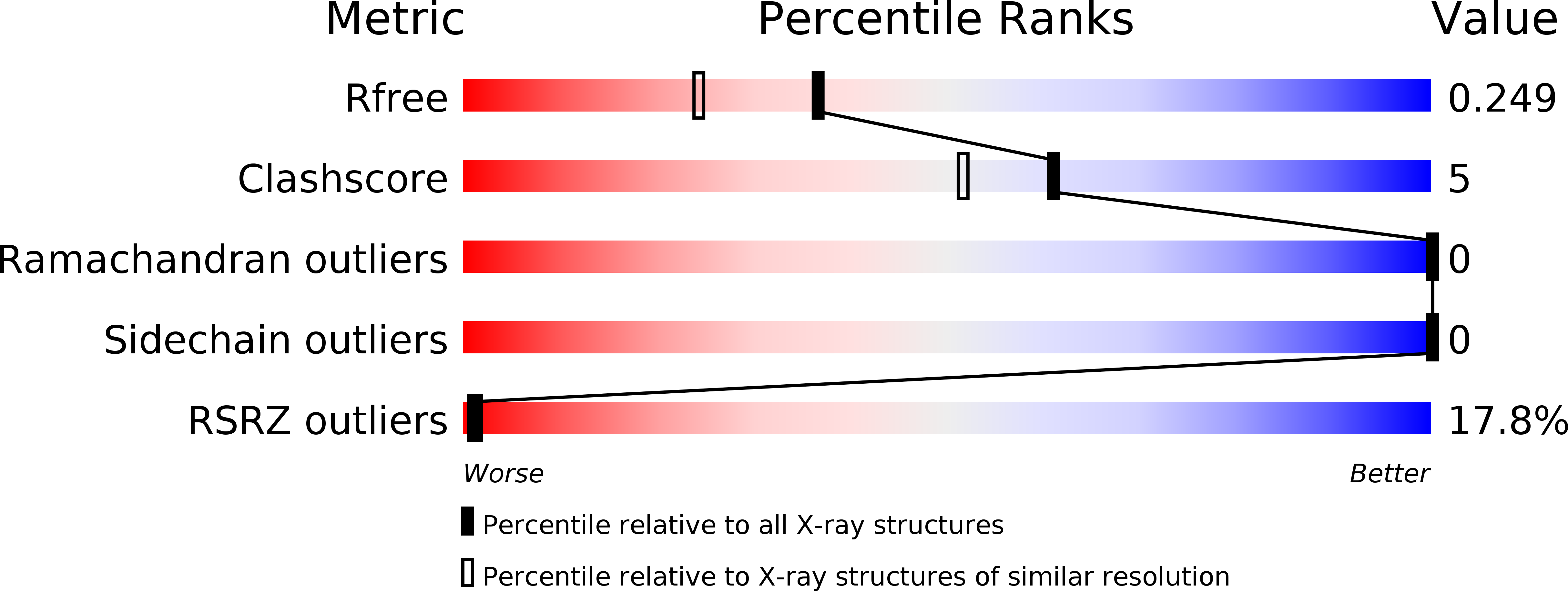
R-Value Free:
0.24
R-Value Work:
0.21
R-Value Observed:
0.21
Space Group:
C 2 2 21