4Q5O image
Deposition Date 2014-04-17
Release Date 2014-09-10
Last Version Date 2023-09-20
Entry Detail
PDB ID:
4Q5O
Keywords:
Title:
Crystal structure of EctD from S. alaskensis with 2-oxoglutarate and 5-hydroxyectoine
Biological Source:
Source Organism:
Host Organism:
Method Details:
Experimental Method:
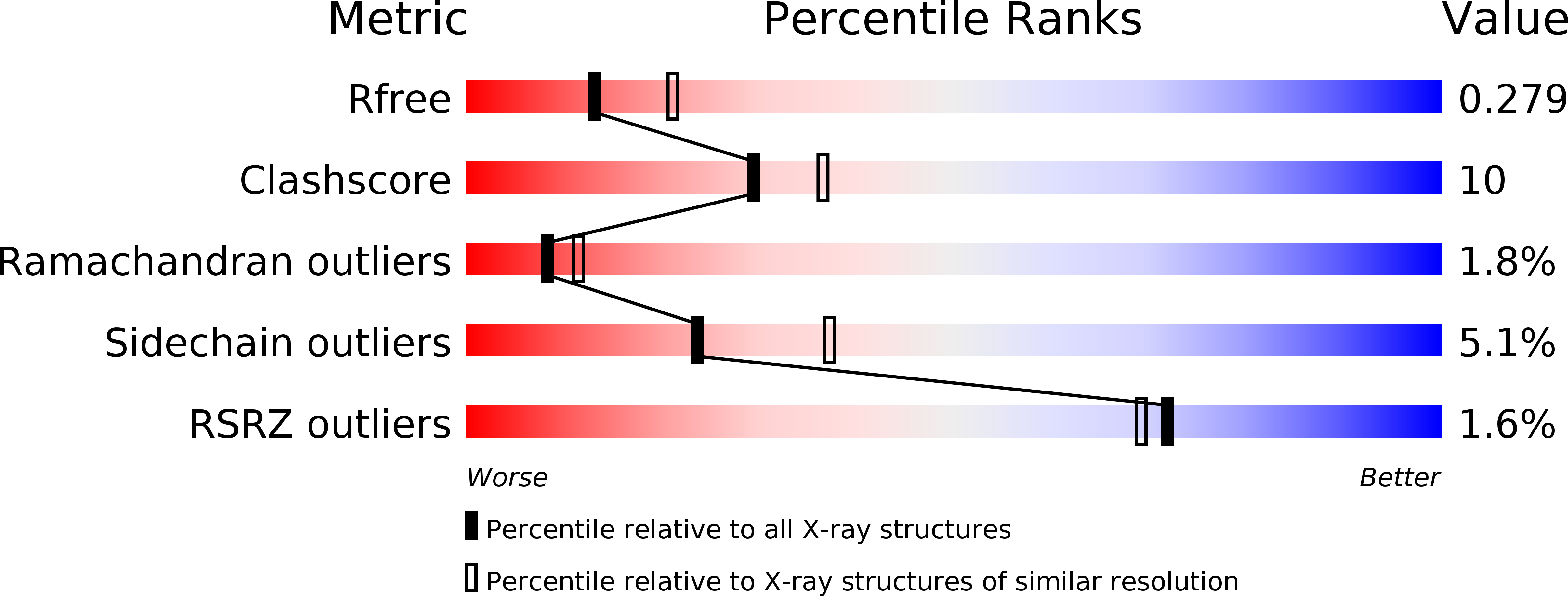
Resolution:
2.64 Å
R-Value Free:
0.27
R-Value Work:
0.20
R-Value Observed:
0.21
Space Group:
P 21 21 21
Macromolecular Entities
Structures with similar UniProt ID
Protein Blast
Polymer Type:polypeptide(L)
Molecule:Ectoine hydroxylase
Gene (Uniprot):ectD
Chain IDs:A, B
Chain Length:314
Number of Molecules:2
Biological Source:Sphingopyxis alaskensis RB2256
Primary Citation
Crystal structure of the ectoine hydroxylase, a snapshot of the active site.
J.Biol.Chem. 289 29570 29583 (2014)
PMID: 25172507 DOI: 10.1074/jbc.M114.576769

Abstact

Ectoine and its derivative 5-hydroxyectoine are compatible solutes that are widely synthesized by bacteria to cope physiologically with osmotic stress. They also serve as chemical chaperones and maintain the functionality of macromolecules. 5-Hydroxyectoine is produced from ectoine through a stereo-specific hydroxylation, an enzymatic reaction catalyzed by the ectoine hydroxylase (EctD). The EctD protein is a member of the non-heme-containing iron(II) and 2-oxoglutarate-dependent dioxygenase superfamily and is evolutionarily well conserved. We studied the ectoine hydroxylase from the cold-adapted marine ultra-microbacterium Sphingopyxis alaskensis (Sa) and found that the purified SaEctD protein is a homodimer in solution. We determined the SaEctD crystal structure in its apo-form, complexed with the iron catalyst, and in a form that contained iron, the co-substrate 2-oxoglutarate, and the reaction product of EctD, 5-hydroxyectoine. The iron and 2-oxoglutarate ligands are bound within the EctD active site in a fashion similar to that found in other members of the dioxygenase superfamily. 5-Hydroxyectoine, however, is coordinated by EctD in manner different from that found in high affinity solute receptor proteins operating in conjunction with microbial import systems for ectoines. Our crystallographic analysis provides a detailed view into the active site of the ectoine hydroxylase and exposes an intricate network of interactions between the enzyme and its ligands that collectively ensure the hydroxylation of the ectoine substrate in a position- and stereo-specific manner.

Legend

Protein

Chemical

Disease

Primary Citation of related structures