4Q56 image
Deposition Date 2014-04-16
Release Date 2015-10-14
Last Version Date 2024-11-06
Entry Detail
PDB ID:
4Q56
Title:
Structure of Helix aspersa agglutinin with natural glycosylation and N-acetyl-alpha-D-galactosamine (GalNAc)
Biological Source:
Source Organism:
Helix aspersa (Taxon ID: 6535)
Method Details:
Experimental Method:
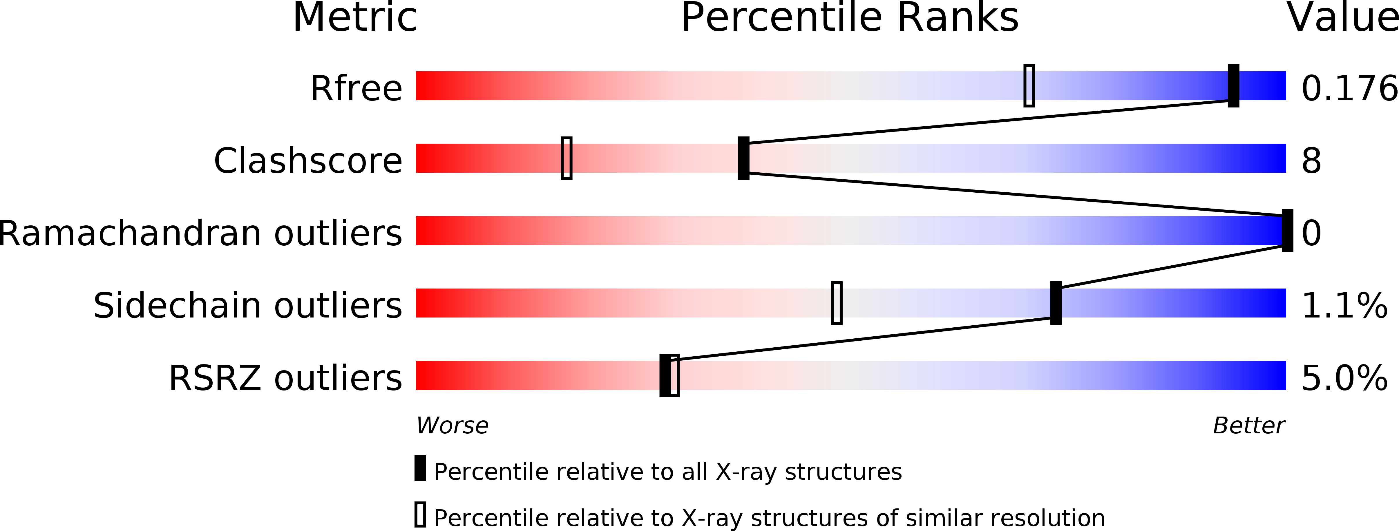
Resolution:
1.38 Å
R-Value Free:
0.16
R-Value Work:
0.13
R-Value Observed:
0.13
Space Group:
H 3 2
Macromolecular Entities
Polymer Type:polypeptide(L)
Molecule:Helix aspersa agglutinin (HAA)
Chain IDs:A
Chain Length:101
Number of Molecules:1
Biological Source:Helix aspersa
Modified Residue
Compound ID Chain ID Parent Comp ID Details 2D Image
ASN A ASN GLYCOSYLATION SITE
PCA A GLN PYROGLUTAMIC ACID
Primary Citation
Structural studies of Helix aspersa agglutinin complexed with GalNAc: A lectin that serves as a diagnostic tool.
Int.J.Biol.Macromol. 81 1059 1068 (2015)
PMID: 26416237 DOI: 10.1016/j.ijbiomac.2015.09.044

Abstact

Lectins belong to a differentiated group of proteins known to possess sugar-binding properties. Due to this fact, they are interesting research targets in medical diagnostics. Helix aspersa agglutinin (HAA) is a lectin that recognizes the epitopes containing α-d-N-acetylgalactosamine (GalNAc), which is present at the surface of metastatic cancer cells. Although several reports have already described the use of HAA as a diagnostic tool, this protein was not characterized on the molecular level. Here, we present for the first time the structural information about lectin isolated from mucus of Helix aspersa (garden snail). The amino acid sequence of this agglutinin was determined by Edman degradation and tertiary as well as quaternary structure by X-ray crystallography. The high resolution crystal structure (1.38Å) and MALDI-TOF mass spectrometry analysis provide the detailed information about a large part of the HAA natural glycan chain. The topology of the GalNAc binding cleft and interaction with lectin are very well defined in the structure and fully confirmed by STD HSQC NMR spectroscopy. Together, this provides structural clues regarding HAA specificity and opens possibilities to rational modifications of this important diagnostic tool.

Legend

Protein

Chemical

Disease

Primary Citation of related structures