Deposition Date
2014-04-10
Release Date
2014-05-07
Last Version Date
2025-03-26
Entry Detail
PDB ID:
4Q31
Keywords:
Title:
The crystal structure of cystathione gamma lyase (CalE6) from Micromonospora echinospora
Biological Source:
Source Organism(s):
Micromonospora echinospora (Taxon ID: 1877)
Expression System(s):
Method Details:
Experimental Method:
Resolution:
2.10 Å
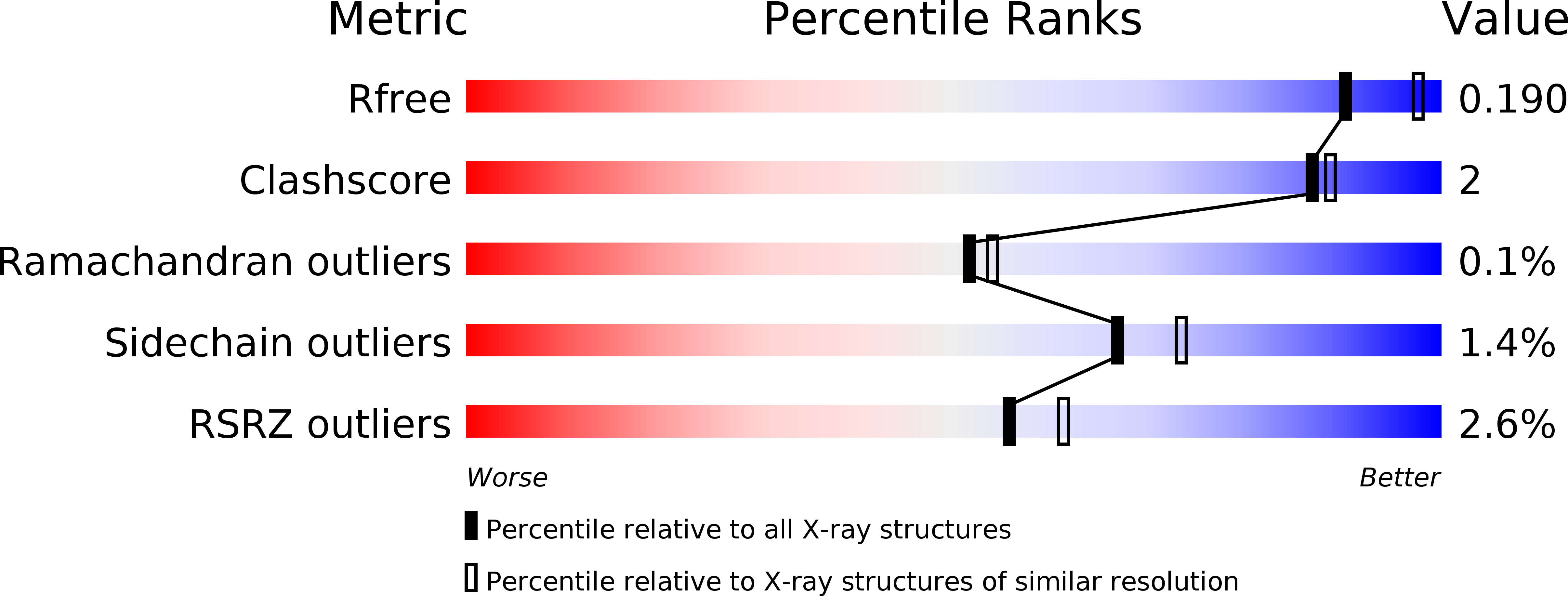
R-Value Free:
0.19
R-Value Work:
0.15
R-Value Observed:
0.15
Space Group:
I 2 2 2