Deposition Date
2014-04-10
Release Date
2014-07-16
Last Version Date
2024-10-30
Entry Detail
PDB ID:
4Q2W
Keywords:
Title:
Crystal Structure of pneumococcal peptidoglycan hydrolase LytB
Biological Source:
Source Organism(s):
Streptococcus pneumoniae (Taxon ID: 170187)
Expression System(s):
Method Details:
Experimental Method:
Resolution:
1.65 Å
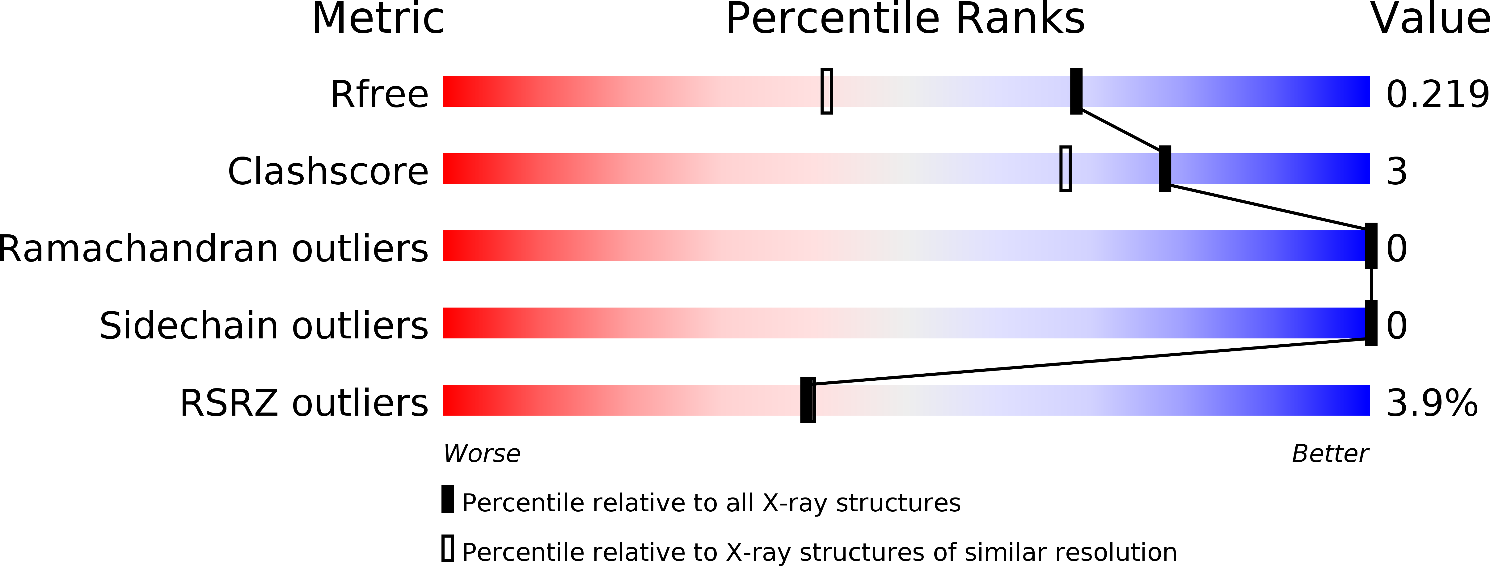
R-Value Free:
0.21
R-Value Work:
0.18
R-Value Observed:
0.18
Space Group:
C 1 2 1