4Q2J image
Deposition Date 2014-04-08
Release Date 2015-03-11
Last Version Date 2024-03-20
Entry Detail
PDB ID:
4Q2J
Title:
A novel structure-based mechanism for DNA-binding of SATB1
Biological Source:
Source Organism:
Mus musculus (Taxon ID: 10090)
Host Organism:
Method Details:
Experimental Method:
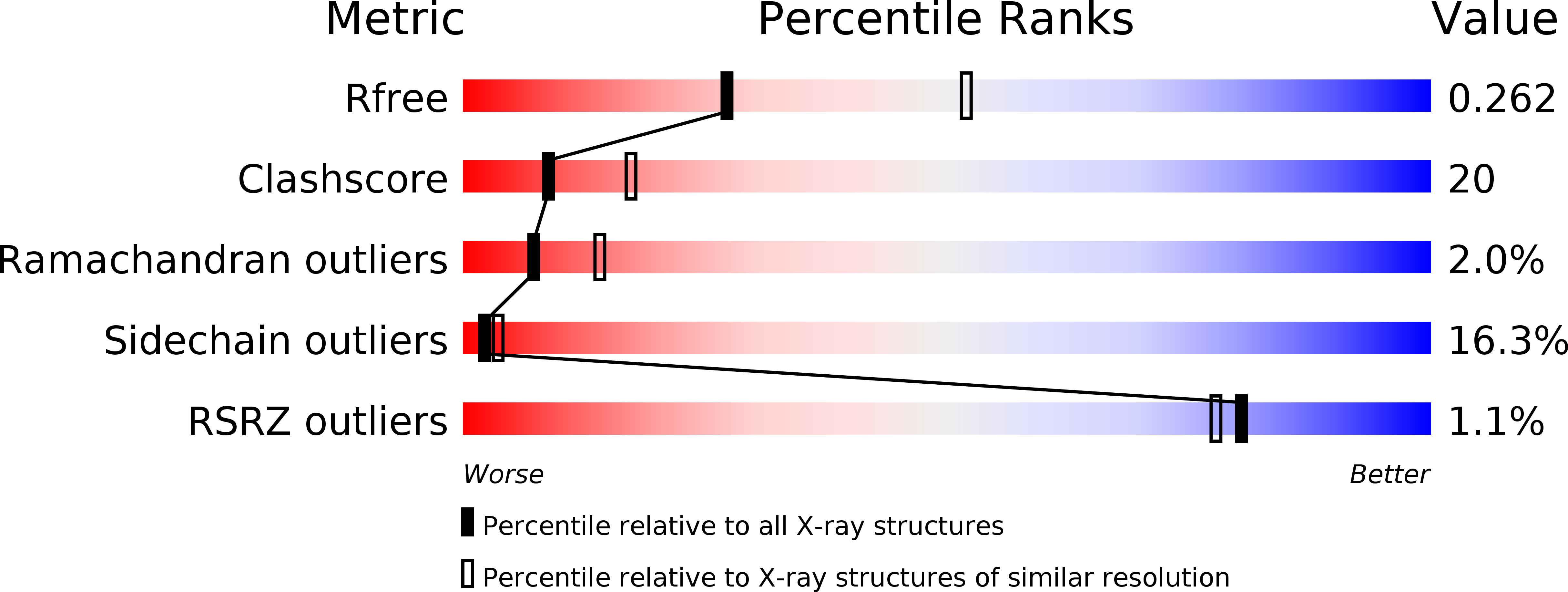
Resolution:
2.60 Å
R-Value Free:
0.27
R-Value Work:
0.22
R-Value Observed:
0.23
Space Group:
C 1 2 1
Macromolecular Entities
Structures with similar UniProt ID
Protein Blast
Polymer Type:polypeptide(L)
Molecule:DNA-binding protein SATB1
Gene (Uniprot):Satb1
Chain IDs:A, B, C, D
Chain Length:180
Number of Molecules:4
Biological Source:Mus musculus
Primary Citation
Crystal structure of the ubiquitin-like domain-CUT repeat-like tandem of special AT-rich sequence binding protein 1 (SATB1) reveals a coordinating DNA-binding mechanism.
J.Biol.Chem. 289 27376 27385 (2014)
PMID: 25124042 DOI: 10.1074/jbc.M114.562314

Abstact

SATB1 is essential for T-cell development and growth and metastasis of multitype tumors and acts as a global chromatin organizer and gene expression regulator. The DNA binding ability of SATB1 plays vital roles in its various biological functions. We report the crystal structure of the N-terminal module of SATB1. Interestingly, this module contains a ubiquitin-like domain (ULD) and a CUT repeat-like (CUTL) domain (ULD-CUTL tandem). Detailed biochemical experiments indicate that the N terminus of SATB1 (residues 1-248, SATB1((1-248))), including the extreme 70 N-terminal amino acids, and the ULD-CUTL tandem bind specifically to DNA targets. Our results show that the DNA binding ability of full-length SATB1 requires the contribution of the CUTL domain, as well as the CUT1-CUT2 tandem domain and the homeodomain. These findings may reveal a multiple-domain-coordinated mechanism whereby SATB1 recognizes DNA targets.

Legend

Protein

Chemical

Disease

Primary Citation of related structures