4Q0P image
Deposition Date 2014-04-02
Release Date 2014-05-28
Last Version Date 2024-04-03
Entry Detail
PDB ID:
4Q0P
Keywords:
Title:
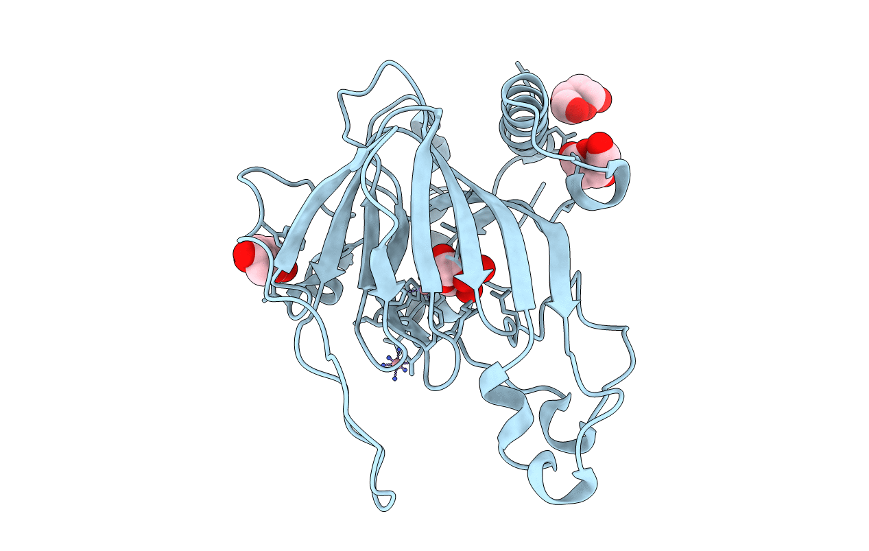
Crystal structure of Acinetobacter sp. DL28 L-ribose isomerase in complex with L-ribose
Biological Source:
Source Organism:
Acinetobacter (Taxon ID: 160971)
Host Organism:
Method Details:
Experimental Method:
Resolution:
1.93 Å
R-Value Free:
0.24
R-Value Work:
0.20
R-Value Observed:
0.20
Space Group:
F 2 2 2
Macromolecular Entities
Structures with similar UniProt ID
Protein Blast
Polymer Type:polypeptide(L)
Molecule:L-Ribose isomerase
Gene (Uniprot):L-RI
Chain IDs:A
Chain Length:260
Number of Molecules:1
Biological Source:Acinetobacter
Primary Citation
X-ray structure of a novel L-ribose isomerase acting on a non-natural sugar L-ribose as its ideal substrate.
Febs J. 281 3150 3164 (2014)
PMID: 24846739 DOI: 10.1111/febs.12850

Abstact

UNLABELLED l-Ribose, a pentose, is not known to exist in nature. Although organisms typically do not have a metabolic pathway that uses l-ribose as a carbon source, prokaryotes use various sugars as carbon sources for survival. Acinetobacter sp. DL-28 has been shown to express the novel enzyme, l-ribose isomerase (AcL-RbI), which catalyzes reversible isomerization between l-ribose and l-ribulose. AcL-RbI showed the highest activity to l-ribose, followed by d-lyxose with 47% activity, and had no significant amino acid sequence similarity to structure-known proteins, except for weak homology with the d-lyxose isomerases from Escherichia coli O157 : H7 (18%) and Bacillus subtilis strain (19%). Thus, AcL-RbI is expected to have the unique three-dimensional structure to recognize l-ribose as its ideal substrate. The X-ray structures of AcL-RbI in complexes with substrates were determined. AcL-RbI had a cupin-type β-barrel structure, and the catalytic site was found between two large β-sheets with a bound metal ion. The catalytic site structures clearly showed that AcL-RbI adopted a cis-enediol intermediate mechanism for the isomerization reaction using two glutamate residues (Glu113 and Glu204) as acid/base catalysts. In its crystal form, AcL-RbI formed a unique homotetramer with many substrate sub-binding sites, which likely facilitated capture of the substrate. DATABASE The atomic coordinates and structure factors of AcL-RbI/l-ribose, AcL-RbI/l-ribulose, AcL-RbI/ribitol, E204Q/l-ribose and E204Q/l-ribulose have been deposited in the Protein Data Bank under accession codes, 4Q0P, 4Q0Q, 4Q0S, 4Q0U and 4Q0V.

Legend

Protein

Chemical

Disease

Primary Citation of related structures
Feedback Form
Name
Email
Institute
Feedback