Deposition Date
2014-03-25
Release Date
2014-04-30
Last Version Date
2024-10-30
Entry Detail
PDB ID:
4PY0
Keywords:
Title:
Crystal structure of P2Y12 receptor in complex with 2MeSATP
Biological Source:
Source Organism(s):
Homo sapiens (Taxon ID: 9606)
Escherichia coli (Taxon ID: 562)
Escherichia coli (Taxon ID: 562)
Expression System(s):
Method Details:
Experimental Method:
Resolution:
3.10 Å
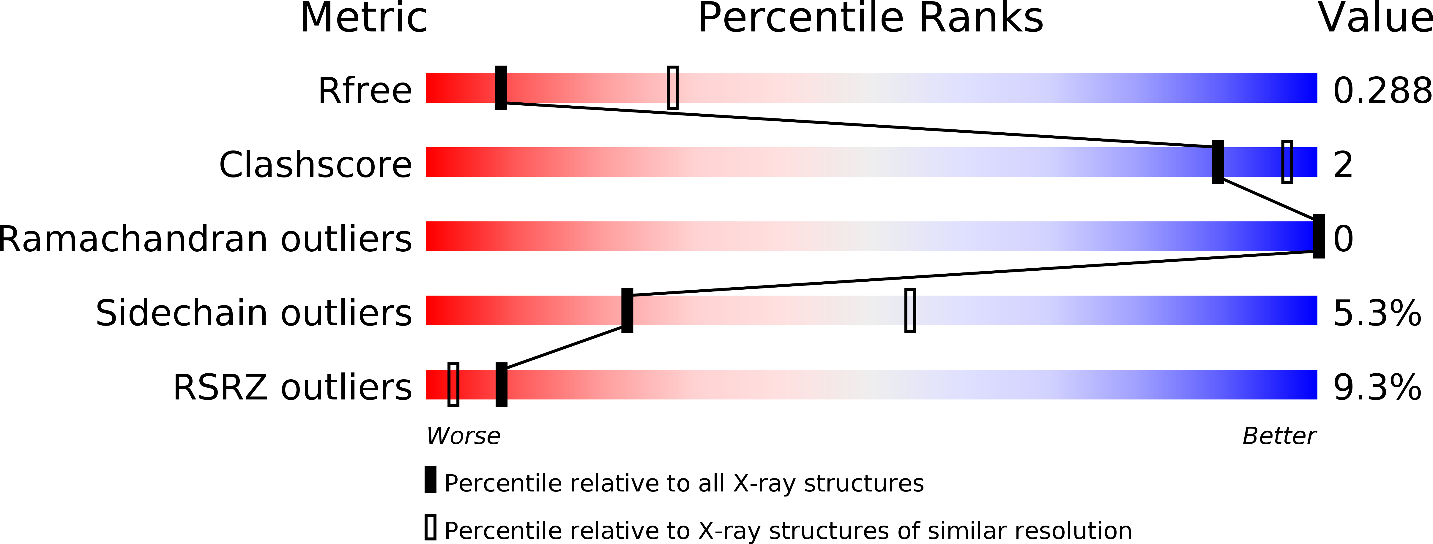
R-Value Free:
0.26
R-Value Work:
0.22
R-Value Observed:
0.22
Space Group:
C 1 2 1