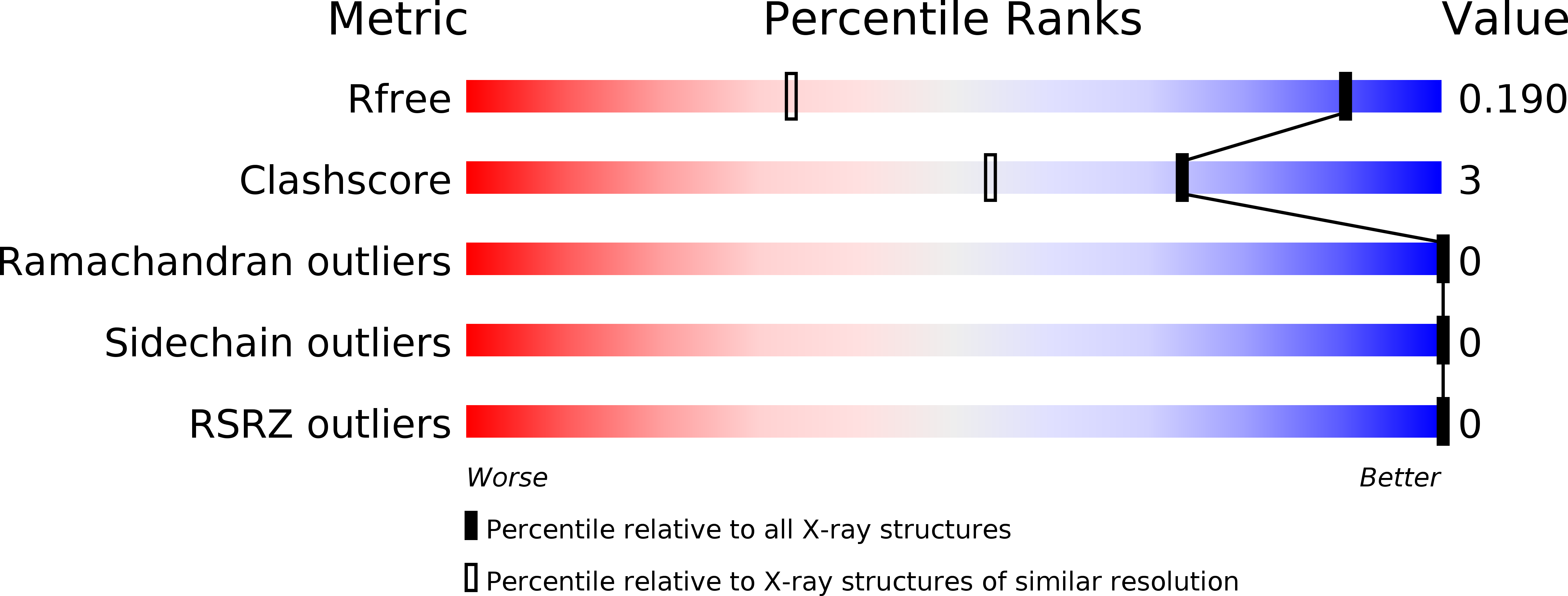
4PX8 image
Deposition Date 2014-03-22
Release Date 2015-10-21
Last Version Date 2023-09-20
Entry Detail
PDB ID:
4PX8
Keywords:
Title:
Structure of P. vulgaris HigB toxin
Biological Source:
Source Organism:
Proteus vulgaris (Taxon ID: 585)
Host Organism:
Method Details:
Experimental Method:
Resolution:
1.25 Å
R-Value Free:
0.18
R-Value Work:
0.15
R-Value Observed:
0.15
Space Group:
C 1 2 1
Macromolecular Entities
Structures with similar UniProt ID
Protein Blast
Polymer Type:polypeptide(L)
Molecule:Killer protein
Gene (Uniprot):higB
Chain IDs:A
Chain Length:118
Number of Molecules:1
Biological Source:Proteus vulgaris
Ligand Molecules
Primary Citation
Defining the mRNA recognition signature of a bacterial toxin protein.
Proc.Natl.Acad.Sci.USA 112 13862 13867 (2015)
PMID: 26508639 DOI: 10.1073/pnas.1512959112

Abstact

Bacteria contain multiple type II toxins that selectively degrade mRNAs bound to the ribosome to regulate translation and growth and facilitate survival during the stringent response. Ribosome-dependent toxins recognize a variety of three-nucleotide codons within the aminoacyl (A) site, but how these endonucleases achieve substrate specificity remains poorly understood. Here, we identify the critical features for how the host inhibition of growth B (HigB) toxin recognizes each of the three A-site nucleotides for cleavage. X-ray crystal structures of HigB bound to two different codons on the ribosome illustrate how HigB uses a microbial RNase-like nucleotide recognition loop to recognize either cytosine or adenosine at the second A-site position. Strikingly, a single HigB residue and 16S rRNA residue C1054 form an adenosine-specific pocket at the third A-site nucleotide, in contrast to how tRNAs decode mRNA. Our results demonstrate that the most important determinant for mRNA cleavage by ribosome-dependent toxins is interaction with the third A-site nucleotide.

Legend

Protein

Chemical

Disease

Primary Citation of related structures